A new functional bone imaging technique that combines PET and MRI shows that bone metabolism is higher in patients with osteoarthritis in the knee, according to research presented July 13 at the Society of Nuclear Medicine and Molecular Imaging (SNMMI) 2020 virtual annual meeting.
The technique offers another way to assess degeneration of the knee joint, a team led by Lauren Watkins of Stanford University's Imaging of Musculoskeletal Function Group said in a statement released by SNMMI.
"[Our] findings show the utility and potential of PET imaging to study the role of bone physiology in degenerative joint disease," Watkins said. "This knowledge may help us understand the order of events leading to structural and functional degeneration of the knee."
Osteoarthritis affects more than 32.5 million adults in the U.S., the SNMMI said. For their study, Watkins and colleagues used 18F-FDG PET with MRI to visualize the knees of 12 study participants. Five of these 12 underwent two scans with at least five days in between to determine whether the technique was effectively repeatable. A musculoskeletal radiologist assessed each knee using the MRI Osteoarthritis Knee Score (MOAKS) and PET data to calculate rates of bone perfusion, tissue mineralization, and the tracer uptake rate in the bone.
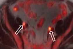
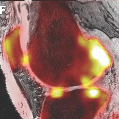
![Representative structural MRI image (proton-density-weighted IDEAL [iterative decomposition of water and fat with echo asymmetry and least-squares estimation] image, A and D), 18F-NaF PET standardized uptake value images (B and E), and PET/MR fusion images (C and F) of a healthy knee and a knee with osteoarthritis. MOAKS scoring of MR images was used to identify the size of osteophytes, bone marrow lesions, and cartilage loss within various bone regions in the patella, tibia, and femur. Arrows highlight bone regions with osteophytes in blue, bone marrow lesions in yellow, and cartilage loss in green. Image courtesy of Lauren Watkins, et al, Stanford University.](https://img.auntminnie.com/files/base/smg/all/image/2020/07/am.2020_07_14_20_06_2345_2020_07_13_SNMMI_bone_imaging_20200714204326.png?auto=format%2Ccompress&dpr=2&fit=max&q=70&w=700) Representative structural MRI image (proton-density-weighted IDEAL [iterative decomposition of water and fat with echo asymmetry and least-squares estimation] image, A and D), 18F-NaF PET standardized uptake value images (B and E), and PET/MR fusion images (C and F) of a healthy knee and a knee with osteoarthritis. MOAKS scoring of MR images was used to identify the size of osteophytes, bone marrow lesions, and cartilage loss within various bone regions in the patella, tibia, and femur. Arrows highlight bone regions with osteophytes in blue, bone marrow lesions in yellow, and cartilage loss in green. Image courtesy of Lauren Watkins, et al, Stanford University.
Representative structural MRI image (proton-density-weighted IDEAL [iterative decomposition of water and fat with echo asymmetry and least-squares estimation] image, A and D), 18F-NaF PET standardized uptake value images (B and E), and PET/MR fusion images (C and F) of a healthy knee and a knee with osteoarthritis. MOAKS scoring of MR images was used to identify the size of osteophytes, bone marrow lesions, and cartilage loss within various bone regions in the patella, tibia, and femur. Arrows highlight bone regions with osteophytes in blue, bone marrow lesions in yellow, and cartilage loss in green. Image courtesy of Lauren Watkins, et al, Stanford University.On PET, the group found abnormal bone metabolism in areas of the knee with bone marrow lesions, osteophytes, and adjacent cartilage lesion; these characteristics were also associated with higher bone perfusion rates on MRI.
"[Our study results could] help us to develop and quickly evaluate new interventions that target specific metabolic pathways to give us the best chance to slow or arrest the onset and progression of osteoarthritis," Watkins said.