The U.S. National Institutes of Health (NIH) Clinical Center in Bethesda, MD, has installed its first photon-counting CT scanner to explore use of the investigational technology in a clinical setting.
The prototype scanner (Somatom Count, Siemens Healthcare) is expected to replicate the image quality of conventional CT scanning at a lower radiation dose, while also providing enhanced views of the body via multienergy imaging, NIH said.
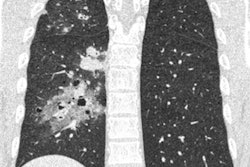
The imaging provided by photon-counting CT will allows clinicians to identify materials such as iodine with great anatomic precision, the agency said. The new technology may help characterize tumors, plaques, and vessels smaller than 0.5 mm, as well as identify soft tissues such as tendons or collagen, which are hard to differentiate with current equipment.
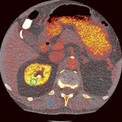
 Photon-counting abdominal CT scan of a patient at the center shows increased iodine uptake in bright yellow areas. Image courtesy of the NIH.
Dr. David Bluemke, PhD, chief of radiology, and colleagues will spend five years developing protocols and processing algorithms aimed at improving screening, imaging, and treatment planning for health conditions ranging from cancer to cardiovascular disease.
"Now is an exciting time for us and for our study participants here in the Clinical Center as we help test and develop this CT technology so that it may one day help patients around the world and impact the healthcare they receive," Bluemke said in a statement.
Through its cooperative research and development agreement with Siemens, the center is testing the technology to help optimize the scanner for clinical use in the U.S. and around the world, NIH said.