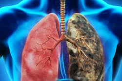
How low can chest CT doses really go? Researchers from Boston may have bested even the chest x-ray, the ultimate low-dose scan, using a special data sampling technique to produce diagnostic-quality CT lung screening scans at doses as low as 0.25 mSv.
The group from Massachusetts General Hospital (MGH) used a technique called sparse data sampling in conjunction with iterative reconstruction to produce readable lung scans in normal adult men at 0.25 mSv. The results not only equaled the accuracy of standard-dose scans reconstructed with filtered back projection algorithms (scans acquired at several times the dose), they were superior to low-dose images reconstructed with filtered back projection.
 Dr. Ranish Khawaja of Massachusetts General Hospital.
"More than 90% dose reduction can be achieved with sparsely sampled data compared to fully sampled data in chest CT for lung evaluation," said Dr. Ranish Khawaja in a presentation at the recent RSNA annual meeting.
The sparse sampling approach
The most common dose-reduction technique is to reduce CT tube current, but doing so increases electronic image noise, and iterative reconstruction can only partially correct for this, he said.
"With a sparse sample approach, we can still have high tube current, but we reduce the projection fields," Khawaja explained. "How does it work? High-dose chest CT projection data are reconstructed in a noncontinuous fashion, with angular gaps between subsequent projections. That results in sparsely sampled CT data."
Sparse data sampling has already been tried in several CT applications, but thoracic CT has not been one of them, he said. Therefore, the study aimed to assess the role of sparsely sampled data for substantial dose reductions in chest CT.
Khawaja, Dr. Mannudeep Kalra, and colleagues at MGH assembled a study population of 10 male patients (mean age, 64; mean body mass index, 27, with a range of 21-32). The individuals were scanned with three techniques:
- Sparsely sampled sub-mSv CT with iterative reconstruction
- Sub-mSv CT with filtered back projection (FBP) reconstruction
- Standard-dose CT with FBP reconstruction
All CT scans were acquired on a 256-detector-row scanner (Brilliance iCT, Philips Healthcare). For the sparsely sampled data technique, the group acquired noncontinuous data with angular gaps per rotation between subsequent projections. Sparse-data scans were acquired at 120 kV, with 0.95 pitch, 2.5-mm section thickness, 8-cm beam width, 128 x 0.625 detector configuration, and tube current modulation.
The sub-mSv exam was performed at identical scan parameters with the exception of tube current, which was manually reduced to 18 mA to 20 mA to obtain chest CT scans at dose-length products of less than 70 mGy-cm and less than 1 mSv, Khawaja said.
"How did we achieve sparse sampling? We discarded double z-sampling first, further discarding every second view to arrive at an effective angular sampling of 600 projections per full 360° turn" of the gantry, he said.
In addition to standard-dose FBP and sub-mSv FBP, sparsely sampled images were generated from raw data by reconstructing 25% of angular projections from sub-mSv sinogram data, then applying a penalized maximum likelihood iterative reconstruction technique, Khawaja said.
Image noise was measured in regions of interest at three anatomic locations. Spectral profiles of noise spectral density were calculated using MatLab software for all image series. Statistical analysis included descriptive statistics, analysis of variance (ANOVA), and the Kappa test for interobserver agreement.
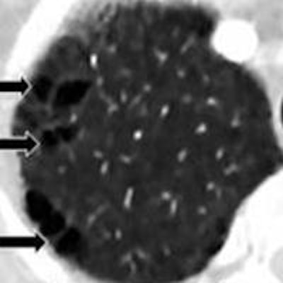
 Chest CT images of a 66-year-old man (body mass index, 21) with emphysema. Margins of air pockets are equal in all three images. No streaking artifacts are present in sparsely sampled sub-mSv with iterative reconstruction, even at 91% dose reduction. Images courtesy of Dr. Ranish Khawaja and Dr. Mannudeep Kalra.
Two experienced radiologists performed independent and blinded evaluations for lesion detection, lesion margins, diagnostic confidence, visibility of small pulmonary structures, artifacts, peripheral lung vessels, and subsegmental bronchi, Khawaja said.
"We did not assess the mediastinal soft tissues; the intent was primarily to assess dose-reduction values," he added.
The results showed "perfect interobserver agreement between the two radiologists," he said. The 46 abnormalities included 33 solid nodules and were seen across all three datasets. Lesion margins in 17 of 20 lesions were deemed "acceptable" with sparsely sampled images.
Noise spectral density measurements showed an exponential decrease of noise power over frequency with sparsely sampled data in a semilog plot, compared with sub-mSv FBP and standard-dose FBP images.
Diagnostic confidence with sparsely sampled data was equivalent to that of standard-dose FBP in nine of 10 patients, the researchers found.
"You can see streak artifacts and a little haziness in the sparsely sampled images," Khawaja said. "Minor pixelation artifacts were seen in three of 10 patients, and one patient had a streak artifact, but they were of no clinical significance."
"CT radiation dose reduction in chest CT is important and possible," he said. Radiologists interpreting scans reconstructed with sparsely sampled data had equal diagnostic confidence to those interpreting full-dose FBP scans in chest CT for lung evaluation. In fact, more than 90% dose reduction can be achieved using sparsely sampled versus fully sampled CT data.
"These findings may have a potential role in lung cancer screening at ultralow radiation dose," Khawaja said.
Future studies will help explain the effect of a sparsely sampled iterative reconstruction technique on mediastinal and abdominal soft tissues, which have lower inherent tissue contrast compared to the lungs, he added.