(Radiology Review) For patients with mammographically occult breast lesions, an additional lesion marker placed at needle localization may enable immediate confirmation of lesion excision, according to radiologists at New York University School of Medicine in New York City.
Dr. Cecilia Mercado and colleagues reviewed the results of 135 ultrasound-guided needle localizations during a 21-month period for a study published in the American Journal of Roentgenology (October 2008, Vol. 191:4, pp. 1216-1219).
Because some breast malignancies are visualized with sonography but not mammography, the authors used sonography for preoperative marker placement to assess whether this might improve the accuracy of lesion excision and adequate inclusion of lesion margins.
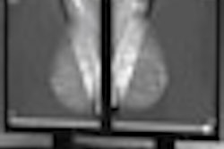
The researchers retrospectively evaluated lesion type and size and appropriate wire and marker placement. Using mammography, they evaluated the distance between the localizing wire and lesion marker. Also, specimen radiographs were assessed for wire and localizing markers within the lesion.
A 21-gauge modified Kopans spring-hook localization needle was placed into and through the lesion under ultrasound guidance. A hookwire was inserted through the needle and the needle was removed. A 14-gauge introducer was used to place the breast marker, which was a localizing clip embedded in a bioresorbable collagen plug (Cormark, Ethicon Endo-Surgery, Cincinnati).
The researchers divided 121 patients into two groups, but only one group had markers placed in the lesion following needle localization. For the group with markers placed, the authors confirmed lesion or marker excision using specimen radiography (at surgery) in 97% of lesions. However, for the group without markers placed, they confirmed lesion excision using specimen radiography in 23% of lesions.
Results showed that "of the 11 malignant lesions (19%) localized with marker placement, none had a positive inked margin, but five (46%) had close margins necessitating re-excision. Of the 26 malignant lesions (34%) localized without marker placement, two (8%) had a positive inked margin, and eight (31%) had close margins necessitating re-excision," they wrote.
The authors concluded that placement of a marker "aids in identifying mammographically occult lesions within specimens and enables immediate confirmation of accurate surgical removal of localized lesions at surgical excision." Although the study demonstrated an increase in the number of lesions with clear inked margins, there was no significant difference in the number of lesions needing re-excision.
"Sonographically Guided Marker Placement for Confirmation of Removal of Mammographically Occult Lesions After Localization"
C. Mercado et al
Department of Radiology, New York University School of Medicine, New York University Cancer Institute, 160 E. 34th St., 3rd Floor, New York, NY 10016
AJR 2008; 191:1216-1219
By Radiology Review
December 30, 2008
Copyright © 2008 AuntMinnie.com