The full potential of molecular breast imaging for detecting malignant tumors has been constrained by different factors. Conventional scintimammography with the radionuclide Tc-99m sestamibi has been limited by the capabilities of the technology to detect malignant breast tumors smaller than 10 mm in size. Breast PET imaging has been restricted to the use of FDG as a radionuclide, which is not as sensitive for some types of breast malignancies.
A group of researchers from the Mayo Clinic in Rochester, MN, may have overcome these drawbacks by developing and deploying a solid-state dual-head gamma camera for molecular breast imaging.
"This dual-head configuration has the advantage of reducing the maximum distance between the lesion and either detector head to half of the total breast thickness, possibly increasing detection of small lesions without additional imaging time or dose," the authors wrote in a poster presentation exhibited at the 2006 RSNA conference in Chicago.
They also noted that studies in which the lesion is visible in both opposing views can be used to perform quantitative analysis of lesion parameters such as size, depth in the breast, and relative radiotracer uptake.
The team modified a mammographic gantry on which they mounted two opposing field-of-view cadmium zinc telluride (CZT) solid-state detector gamma cameras. The initial configuration used a GE Healthcare (Chalfont St. Giles, U.K.) prototype CZT detector on the upper head and a Gamma Medica-Ideas (Northridge, CA) LumaGem 3200S CZT detector on the lower head. According to the authors, these detectors had intrinsic resolutions of 2.5 mm and 1.6 mm, respectively. More recent studies conducted by the scientists have paired only GE or only Gamma Medica-Ideas detectors on the upper and lower heads.
The dual-head configuration allowed the researchers to image the inferior and lateral aspects of the breast with the lower detector, and the superior and medial breast with the upper detector.
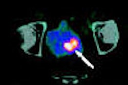
For the study, the team selected patients who had a suspicious lesion (BI-RAD 4-5) that was identified by mammography or ultrasound as smaller than 2 cm in diameter and were scheduled for biopsy. They reported that the study cohort received 20 mCi of Tc-99m sestamibi, with each breast imaged with the dual-head system in craniocaudal (CC) and mediolateral oblique (MLO) positions for 10 minutes per view.
The patients underwent light compression between the detector heads during the imaging procedure, which was conducted while they were seated. All images were read by the same radiologist, and all positive lesions were evaluated for detectability in each of the four views -- lower CC, upper CC, lower MLO, and upper MLO -- acquired by the system.
There were 100 patients participating in the study, with 82 lesions confirmed as cancer in 54 patients. The dual-head system detected 76 of these cancers for an overall sensitivity of 93%.
"Of the six false-negative cancers, two were missed due to inadequate positioning of the patient, and three were less than 5 mm in size," the authors wrote. "Seven additional cancers that were occult on mammography and ultrasound were found with molecular breast imaging."
Sensitivity was affected by tumor size. For tumors 16-20 mm or larger, the system produced 100% sensitivity. Tumors 11-15 mm demonstrated 95% sensitivity with the equipment, while tumors 6-10 mm recorded 93% sensitivity on the technology. The 12 tumors smaller than 5 mm were true positive in nine instances and false negative in three, for a sensitivity of 75%.
Of note, the sensitivity of the technology and technique did not appear to be tumor-dependent, as the system correctly identified invasive ductal carcinoma, ductal carcinoma in situ, invasive lobular carcinoma, tubular carcinoma, medullary carcinoma, invasive ductal carcinoma/ductal carcinoma in situ, and invasive ductal carcinoma/lobular carcinoma.
The scientists compared the results of their study with conventional scintimammography and a single-head CZT gamma camera system. They observed that all three techniques perform similarly for detecting tumors larger than 15 mm, but that at smaller tumor diameters, the sensitivity for both scintimammography and a single-head system drop off more rapidly than the sensitivity for a dual-head molecular breast imaging system.
The team noted that the radiation exposure to the breast is equivalent to a mammogram, but there is a higher dose to the gastrointestinal tract, as well as to other internal organs.
The Mayo Clinic scientists are excited by the results of their initial investigation and have expanded the scope of molecular breast imaging studies with the system.
The technology is currently under evaluation in a 2,000-patient study to compare the dual-head system with mammography for screening women at high risk of breast cancer who have mammographically dense breasts. At the time of the RSNA presentation, the group had enrolled 260 patients in the trial and had detected four tumors and one atypical ductal hyperplasia with the molecular breast imaging system, compared with one tumor that was detected on mammography.
"Additional benefits of the dual-head system, such as quantitative lesion analysis, are currently under investigation," they wrote.
By Jonathan S. Batchelor
AuntMinnie.com staff writer
December 19, 2006
Related Reading
Whole-body FDG-PET of little use in breast cancer staging, November 26, 2006
Preop FDG-PET spots axillary involvement in breast cancer, may obviate biopsy, August 21, 2006
Breast cancer-involved sentinel nodes often contain micrometastases, August 15, 2006
SLN procedure in pregnant breast cancer patients leads to negligible fetal dose, August 14, 2006
Pinhole technique abets dedicated breast SPECT imaging, April 24, 2003
Copyright © 2006 AuntMinnie.com