Computer-aided detection (CAD) technology with an interval change classifier can improve the accuracy of radiologists in differentiating between benign and malignant breast lesions, according to an article published online before print in Radiology. An interval change classifier utilizes change information extracted from prior and current mammograms and estimates a malignancy rating.
"CAD involving interval change analysis of preselected regions of interest can significantly improve radiologists' accuracy in classifying masses on digitized (film-screen) mammograms as malignant or benign," according to a research team from the University of Michigan Medical Center in Ann Arbor and the U.S. Food and Drug Administration's Center for Devices and Radiological Health (CDRH).
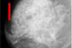
To retrospectively evaluate the effects of CAD involving an interval change classifier, the study team retrospectively reviewed 68 patient files (Radiology, June 26, 2006). Temporal pairs of two-view serial mammograms were obtained from these files and digitized. The mammograms showed 47 malignant and 43 benign biopsy-proven masses.
Eight MQSA-accredited radiologists and two breast imaging fellows then assessed the digitized two-view temporal pairs (in pre-selected regions of interest only) by estimating the likelihood of malignancy and BI-RADS category with and without CAD.
The researchers found that the area under the receiver operating characteristic (ROC) curve for likelihood of malignancy was 0.83 across the 10 observers (range, 0.74-0.88) without CAD and 0.87 (range, 0.80-0.92) with CAD (p < 0.05). The average partial area index above a sensitivity of 0.90 for likelihood of malignancy was 0.35 (range, 0.13-0.54) without CAD and 0.49 (range, 0.18-0.73) with CAD, a difference that failed to reach statistical significance (p = 0.11).
As for BI-RADS assessment, the researchers estimated that six radiologists would have correctly recommended additional biopsies for malignant masses when using CAD (range, 4.3%-10.6%), while five would have correctly recommended fewer biopsies for benign masses (range, 2.3%-9.3%).
The authors also found, however, that five radiologists would have incorrectly recommended additional biopsy for benign masses (range, 2.3%-14%). One would have incorrectly recommended reduction of biopsy (4.3%).
"Our results demonstrate that at both the two-view and the single-view readings there was an improvement in the radiologists' performance when they were assisted by a computer classifier that had a performance in the range that we had studied," the authors concluded.
The researchers acknowledged several limitations in the study, including its use of regions of interest rather than the whole breast and the nonuse of BI-RADS category zero by readers.
"Large prospective clinical trials will be needed to evaluate the effect of CAD on radiologists' diagnostic decisions in clinical settings," the authors stated.
By Erik L. Ridley
AuntMinnie.com staff writer
July 7, 2006
Related Reading
Breast MR falls short for microcalcifications, but CAD helps in malignancies, July 3, 2006
Breast CAD takes aim at architectural distortion, June 30, 2006
Breast CAD put to the test in high-volume community practice, June 26, 2006
CAD finds breast cancers that radiologists missed for years, March 6, 2006
CAD may cut down on missed breast cancers, January 30, 2006
Copyright © 2006 AuntMinnie.com