SAN FRANCISCO - Takotsubo cardiomyopathy accounts for 1% of all myocardial infarction cases in the Japanese population. For some time after its discovery by physicians in 1991, it remained a specifically Japanese syndrome -- until now.
Cases of this transient left ventricular apical ballooning are beginning to pop up in the U.S. Three poster presentations at the American College of Physicians (ACP) meeting focused on how imaging is being used to take a closer look at this condition, named after the Japanese fishing pot designed to catch octopus.
Older women are most susceptible to takotsubo cardiomyopathy in high-pressure situations. That fact was driven home in a case study presented by Drs. Ann Narmi and Amy Arouni from Creighton University Medical Center in Omaha, NE.
"(Takotsubo) is a relatively rare disorder.... Many cases occur in elderly females in a situation of great emotional or physical stress," they wrote.
Their 62-year-old white female presented with shortness of breath, ST-segment changes, and diaphoresis. She had recently witnessed the sudden cardiac death of her daughter. Chest x-ray revealed a diffuse pulmonary edema, while an echocardiogram showed an ejection fraction (EF) of 10% to 15% with global hypokinesis to akinesis.
In a second study, a group from Bridgeport Hospital at Yale University Medical Center in New Haven, CT, described takotsubo as "an enigmatic condition associated with extensive apical and adjacent wall asynergy in a multivessel distribution in the absence of obvious coronary artery disease."
The researchers used myocardial perfusion echocardiography (MPE) in four patients to identify viable myocardium with intact microcirculation. Their patients first underwent coronary angiography, which illustrated nonobstructive coronary artery disease and wall-motion abnormalities.
Myocardial perfusion echo was performed 24 hours after admission and again at 72 hours. The group described one patient as a 67-year-old female who presented with hypercholesterolemia and chest pain. The echo revealed the left ventricle (LV) dilated distally with severe hypokinesis of the mid and apical portion of the LV. The EF estimate was 25%.
"MPE initially showed an absence of perfusion in the apical and adjacent segments," the authors wrote. A repeat MPE at 72 hours indicated remarkable improvement in perfusion, but only a marginal improvement in wall motion.
The group recommended incorporating MPE into the workup of suspected takotsubo cardiomyopathy as a noninvasive way to identify microcirculatory problems that may otherwise be overlooked. This presentation was named a winner in the ACP Vignette Poster Competition.
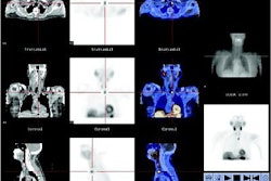
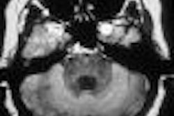
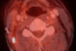
Finally, Dr. Preety Chawla and colleagues from Beth Israel Medical Center in New York City used PET imaging to investigate their case of takotsubo syndrome. Their patient was a 58-year-old woman with a past history of panhypopituitarism, epilepsy, and hyperlipidemia. She presented with severe substernal chest pain and palpitations. She also complained about stress due to family and marital problems.
The ECG showed acute, inferior, ST-elevation myocardial infarction. Coronary angiography revealed normal coronary arteries but reduced LV function with apical akinesis and hyperkinesis of the base. FDG-PET and rubidium-82 PET studies were performed, showing mildly reduced perfusion in the apical region and severely diminished FDG uptake.
"This pattern of relatively normal perfusion with decreased FDG uptake is consistent with viable myocardium," the group wrote. The patient underwent treatment with beta blockers and ACE inhibitors. "At three months, an echocardiogram showed recovered apical function. A repeat PET scan showed markedly improved FDG uptake.... This report ... suggests that (takotsubo) syndrome most likely represents myocardial stunning."
By Shalmali Pal
AuntMinnie.com staff writer
April 19, 2005
Related Reading
Normal angiogram in acute coronary syndrome may warrant other tests, March 29, 2005
Low-dose 40-slice CT sees nonviable myocardium, March 7, 2005
Copyright © 2005 AuntMinnie.com