MR elastography is a potentially useful tool for the detection of malignant breast disease. The technique may also characterize breast lesions more precisely, particularly in women with radiographically dense breasts, according to a study in the American Journal of Roentgenology.
Dr. Alexia McKnight and colleagues at the Mayo Clinic in Rochester, MN, wrote about their experience with an experimental MRI-based method for quantitatively imaging the elastic properties of breast tissue in vivo.
"We wondered which currently unexploited tissue properties might be most useful for detecting and characterizing breast cancer if they could be interrogated with an imaging technique...the elastic modulus of breast carcinoma specimens has been reported to be five-to-20-fold higher than that of adipose-glandular tissue and fibroadenoma specimens, using laboratory mechanical testing," McKnight said (AJR, June 2002, Vol.178:6, pp. 1411-1417).
For this study, the group tested the MR elastography method in phantom breast studies, breast tumor specimens, healthy volunteers, and patients with known breast cancer. The technique relies on vibrational stress to generate shear waves. The minute cyclic displacements associated with shear waves are imaged with motion-sensitive MRI.
An electromechanical driver that is controlled by MR-generated trigger pulses generated shear waves in the object, with displacement amplitudes of less than 500 μm. The MR scan of the propagating waves is processed to create a quantitative map of shear stiffness in the imaging planes.
"The resulting images are equivalent to snapshots of the wave field at a particular phase in the wave cycle," the authors explained. "Typically, four- to eight-wave images are obtained, spanning the wave cycle."
Volunteers and patients were scanned in a prone position, with the breast positioned between two contact plates. Light compression was applied for stabilization. The volunteers did not report any discomfort from the shear-wave driver device, the authors said.
The MR elastograms were performed on a Signa 1.5-tesla scanner (GE Medical Systems, Waukesha, WI). Shear-wave imaging was done with a 2-D gradient-echo MR elastography sequence, in the axial plane, with 3-5-mm section thickness, 64 phase-encoding views, 12-16-cm field of view, a TR range/TE of 100-300/28, and a flip angle of 30° -45° .
Shear-wave frequencies of 75-300 Hz were used for all studies; each wave image required 13-40 seconds for acquisition. Finally, a corresponding T1-weighted image was acquired for anatomic mapping.
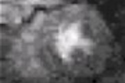
Three tissue-simulating breast phantoms -- one soft, one stiff, and one with a nodule composed of stiffer material -- were constructed to evaluate the driver and radiofrequency coil device. The results of MR elastography in these phantoms showed that the wavelength of the propagating shear wave is longer in the stiff nodule than in the softer surrounding material. "The elastogram depicts a focal area of higher shear stiffness at the location of the hard nodule," the investigators wrote.
Fresh tissue specimens obtained at mastectomy from three patients with invasive ductal carcinoma also were imaged. The MR elastographic scans revealed focal areas of high shear stiffness, corresponding to the location of the palpable tumor. The mean shear stiffness of the three tumors was 580% higher than that of surrounding adipose-fibroglandular tissue, they reported.
Finally, the method was tested in six healthy women and six breast cancer patients, with either infiltrating ductal carcinoma or infiltrating lobular carcinoma. The latter had biopsy-proven palpable breast malignancies of at least 2 cm in diameter.
According to these results, the mean shear stiffness of breast carcinoma was 418% higher than the mean value of surrounding breast tissues. The mean shear stiffness of the tumors was 33 kPa, while the adipose breast tissue measured 8 kPa. The difference in the mean values was statistically significant (p<0.05), according to McKnight and colleagues.
"We speculate that an MR imaging-based technique for quantitatively interrogating the mechanical properties of breast tissue might have potential to increase diagnostic specificity in contrast-enhanced MR imaging examinations that reveal lesions," they wrote.
However, the group said it plans to work on improving the "sharpness and fidelity of MR elastography, thereby potentially further increasing the contrast of small lesions."
The exact properties of the breast that are behind mechanical stiffness are still under investigation as well. At the 2002 American Association of Physicists in Medicine (AAPM) conference in Montreal this month, researchers from Dartmouth-Hitchcock Medical Center and Dartmouth College in Lebanon, NH, presented their work on what they call the "surprisingly complex" nature of breast stiffness.
The team found that some normal, noncancerous breast tissue might be anisotropic. Thus, the shear force may depend on which direction the tissue is pushed or pulled. In studies of two human subjects, the researchers found most tissue within the breasts to be isotropic, but they also detected regions that were clearly anisotropic. As a result, the presence of anisotropic tissue per se is not indicative of breast cancer, the group suggested.
By Shalmali PalAuntMinnie.com staff writer
July 25, 2002
Related Reading
Will breast MRI increase mastectomy rates?, June 28, 2002
MRI successfully tracks women with hereditary breast cancer risk, April 8, 2002
Copyright © 2002 AuntMinnie.com