SEATTLE - If you're looking for the posterior echo patterns of breast nodules, go with pulse-inversion harmonic ultrasound. If it’s the margin and internal echotexture of the nodule that you're after, compound harmonic imaging is the better bet. That’s the conclusion reached by investigators from South Korea, who did a comparison study of the two sonographic techniques.
Dr. Bo Kyoung Seo from Korea University Anam Hospital in Seoul presented the results of her group’s study at the American Roentgen Ray Society meeting on Monday. They compared conventional ultrasound with pulse-inversion harmonic sonography and SonoCT real-time compound imaging.
SonoCT (ATL, Bothell, WA) is an "ultrasound technique that uses electronic beam steering of a transducer array to rapidly acquire several (3-9) overlapping scans of an object from different view angles. These single-angle scans are averaged to form a multiangle compound image that is updated in real-time with each subsequent scan. Compound imaging shows improved image quality compared with conventional ultrasound, primarily because of reduction of speckle, clutter, and other acoustic artifacts," (Seminars in Ultrasound, CT, & MR, February 2001, Vol.22:1, pp.50-64).
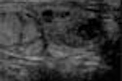
For the Korea University study, images from the same section of 52 breast nodules were taken with all three techniques, using the HDI 5000 (broad bandwidth, 12-5 MHz; linear array, 50 mm). The points of evaluation were contrast between nodule and normal breast; margin; internal echotexture; and posterior echo pattern of the nodules.
The images were assessed by three radiologists and graded on a scale of 1 to 3. Intraobserver agreement in image interpretation was calculated with kappa statistics and Kendall coefficients.
SonoCT was the best technique for evaluation of contrast, margin, and internal echotexture of the nodules (p<0.05), Seo reported. Pulse-inversion harmonic imaging was better suited for evaluation of posterior echo patterns of the nodule (p<0.05). Both of the enhanced techniques were better than conventional sonography all around, Seo said.
One audience member asked how difficult it was to do all three techniques on one patient.
"We use one machine for all three modalities. The time interval between patients is very short," she explained. "There is one switch to change modalities."
In the same session, researchers from Mount Sinai Hospital in Toronto reported similar positive results with SonoCT compound imaging. In one study, they assessed whether compound imaging improved margin resolution and perilesional tissue visualization of solid breast nodules.
One hundred image pairs of solid breast nodules that had been obtained with conventional sonography and SonoCT were reviewed by two blinded breast sonographers. The SonoCT images showed improved margin resolution, perilesional tissue resolution, and retrolesional tissue visualization. Because SonoCT was able to reduce posterior nodule artifact, lesion category was changed from malignant to indeterminate in 7% of the nodules.
In a second study, the Canadian group found that SonoCT improved the imaging of retroareolar tissue, an area that can be difficult to view sonographically because of shadowing. Again, two blinded breast sonographers compared SonoCT images with conventional ultrasound in the retroareolar region of 40 patients.
According to the results, SonoCT images showed grade improvements in parenchymal tissue resolution and a reduction in the density of shadowing.
By Shalmali Pal
AuntMinnie.com staff writer
May 1, 2001
Click here to post your comments about this story. Please include the headline of the article in your message.
Copyright © 2001 AuntMinnie.com