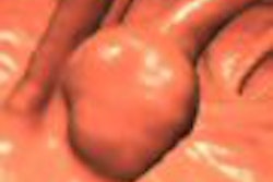
Korean researchers had a hard time finding flat polyps on contrast-enhanced virtual colonoscopy, even with the aid of thin-section imaging on a 16-slice CT scanner, according to a new study. Fewer than half of the flat lesions in the retrospective patient cohort were detected by blinded and unblinded review together.
At the same time, the authors noted that bowel preparation was often suboptimal due to confirmed colon cancer in all the patients. Perhaps more important, fluid and fecal tagging was not used in the study, although many providers consider it essential for optimal visualization, particularly of flat lesions.
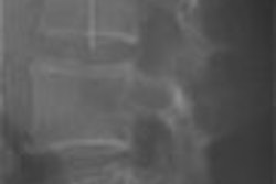
The study, from the Ulsan College of Medicine and Asan Medical Center in Seoul, South Korea, sought to evaluate the ability of contrast-enhanced, 16-slice multidetector-row CT (MDCT) colonography to display flat colonic lesions using a very narrow slice thickness (1 mm).
"Although CT colonography (or virtual colonoscopy, VC) appears to have been successful in extending its role for detecting colonic lesions, there are still some factors that impair lesion detection," wrote Drs. Seong Ho Park, Hyun Kwon Ha, Ah Young Kim, and colleagues (American Journal of Roentgenology, June 2006, Vol. 186:6, pp. 1611-1617).
"Among these factors, flat morphology of a colonic lesion may be an important issue. Despite some controversy ... two large series showed that approximately 38% of the adenomatous lesions detected on colonoscopy were flat," they wrote. "Also, those studies did not clearly address the lesion height, which we hypothesized would be an important factor for determining the visibility of flat colonic lesions."
The cohort consisted of 208 patients, including 180 who were referred for newly diagnosed colon cancer, 20 suspected of having colon cancer, five referred for routine screening, and eight examined for other reasons. The patients had undergone optical colonoscopy prior to CT.
The evening before imaging, patients underwent a cathartic bowel preparation consisting of an oral magnesium carbonate solution (Magcorol Soln, Taejoon Pharmaceuticals, Seoul, South Korea) and 10 mg of bisacodyl laxative the evening before imaging. On the morning of the exam, they were injected with 20 mg of hyoscine N-butylbromide (Buscopan, Boehringer Ingelheim, Ingelheim, Germany) prior to manual room-air insufflation.
Following IV administration of 150 mL iopromide contrast media (Ultravist 370, Schering, Berlin), CT data were acquired on a multidetector scanner (Somatom Sensation 16, Siemens Medical Solutions, Malvern, PA) at 16 x 0.75-mm collimation, 1-mm slice thickness, 0.7-mm reconstruction interval, beam pitch of 1, 120 kVp and 150 mAs.
A board-certified gastrointestinal radiologist with experience in more than 100 VC cases read the data on a dedicated workstation using a software package with volume-rendering capabilities (V-works 5.0, CyberMed, Seoul, South Korea). The reader was blinded to colonoscopy findings and asked to report flat lesions, defined as those with a height less than half their width.
"Lesion size, including the height of the flat lesion, was determined by comparing the lesion with open endoscopic biopsy forceps pushed against the lesion or by direct measure of the resected specimen with a ruler when the lesion was retrieved in toto," the group wrote. Colonoscopy before VC served as the reference standard.
Of 18 flat lesions found at colonoscopy, only four (22.2%, CI 95%) were detected at the blind VC review. When colonoscopy results were revealed for the unblinded review, an additional 7 x 2-mm hyperplastic polyp was found on the CT data. Six lesions could not be visualized even retrospectively because of excessive luminal fluid, poor bowel preparation, and/or poor bowel distension, the authors wrote. And the remaining seven lesions were not visualized despite optimal preparation and distension of the relevant bowel segments.
As for cancers, a 15 x 1-mm adenocarcinoma in situ was not detected at VC due to excessive luminal fluid and poor bowel distension; however two other cancerous lesions were detected at the blinded review, according to the authors.
Lesions had to be 2 mm or greater in height and 7 mm or greater in diameter before they could be detected at VC, the authors concluded, adding that contrast enhancement, location on a haustral fold, and abnormal 2D and D morphology contributed to lesion conspicuity. Additional review with an intermediate soft-tissue window setting after the standard colon window setting did not reveal any additional flat lesions.
"The difficulty of diagnosing flat lesions on CT colonography is also thought to be related to several ... factors," the team wrote. "Plaque-like morphology may be easily mistaken for feces. As shown in the three flat lesions of our study that were confidently distinguished from feces with the help of contrast enhancement, IV contrast may improve interpreter confidence and lesion detection. IV contrast may be used as a problem-solving tool when a potential flat lesion is found and there is doubt; however, the use of the fecal tagging with an oral contrast agent may be a more feasible way to distinguish flat lesions from feces."
In another potential limitation, most patients had advanced colon cancer, which prevented adequate bowel preparation. As a result, many lesions could not be detected due poor bowel preparation and/or excessive luminal fluid.
"Regarding the negative impact of missed flat lesions on screening CT colonography, a recent report suggested that flat lesions were not a significant drawback for screening in an asymptomatic low-risk population," Park and colleagues wrote. "According to some investigators (Pickhardt et al, Radiology 2004; 232:784-790), hyperplastic polyps accounted for most flat lesions in a low-risk screening population. It is also not yet known how many flat lesions in a screening population would be 1 mm or less in height. The scale of the negative impact of missed flat lesions on screening colonography may need further study."
By Eric Barnes
AuntMinnie.com staff writer
June 12, 2006
Related Reading
Prepless VC misses some diminutive flat lesions, January 17, 2006
Telltale barium coating could help VC find risky polyps, March 15, 2006
Virtual colonoscopy picks up flat lesions, November 15, 2004
Virtual colonoscopy aces first flat-lesion study, December 3, 2002
Copyright © 2006 AuntMinnie.com