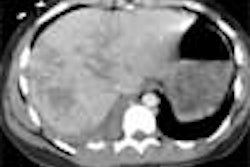
CHICAGO - Finding extracolonic abnormalities is a valuable and documented benefit of virtual colonoscopy, and several studies have shown that clinically significant findings can be followed up at a reasonable cost.
However, researchers have hypothesized that the lack of iodinated contrast in most VC screening exams, as well as the increased focus on low-dose CT protocols, may hinder CT's ability to detect problems outside the colon. There has been little data from which the performance of low-dose screening VC might be compared with its diagnostic counterpart, including their relative accuracy for the detection of extracolonic pathology.
At the RSNA gastrointestinal sessions last week, Dr. Michael Zalis from Massachusetts General Hospital in Boston sought to illuminate the subject with three years of imaging data (2001-2004). The retrospective study examined 650 patients who underwent both screening and diagnostic follow-up VC within six months at MGH. A key question is whether low-dose VC misses important pathology that diagnostic CTC finds.
"It's important for us to be able to try to estimate the likelihood of diagnostic errors, permitting us to calibrate, for example, reader thresholds for making extracolonic diagnoses, and as a component of cost-effectiveness analysis at CT colonography," Zalis said. "The purpose of our study was to estimate the likelihood and significance of extracolonic interpretive errors at (VC), making comparison to follow-up diagnostic (VC)."
The retrospective study included 650 patients over three years who underwent screening VC on multidetector-row CT followed by diagnostic VC exams within six months of the initial exam. Two fellowship-trained abdominal radiologists with VC experience in more than 100 cases each read the screening and follow-up VC exams for each patient to assess any diagnostic discrepancies between the two sets of data.
Discrepancies in extracolonic findings were classified into four categories:
- Normal anatomic variants (such as retroaortic renal vein) with no clinical significance
- Lesions deemed to have little or no clinical significance (such as a hepatic cyst)
- Indeterminate lesions for which one might consider further workup
- Findings deemed likely to be significant, for which diagnostic and therapeutic workup were recommended (such as AAA).
Discrepant findings were confirmed by re-evaluation of the relevant data.
Findings appropriately described in both exams were deemed matches, he said. Those that were excluded from the field-of-view on screening VC but detected on diagnostic VC were not counted as VC errors. Abnormalities that resolved or arose de novo within the six-month study window between exams were counted separately, and the group also analyzed errors attributable to the lack of intravenous contrast on the screening exam. Most patients who underwent both exams were symptomatic, and intravenous contrast was used in 25% of the diagnostic VC exams. Four of the follow-up exams were performed due to a call resulting from screening VC, Zalis said.
The results showed 29 extracolonic findings detected at screening VC and/or follow-up. Of these, 14/29 (48%) were matches on both exams, 3/29 (10%) were deemed VC errors in perception or diagnosis, and 12/29 (41%) arose or resolved themselves between exams.
Diagnostic VC detected 149 extracolonic findings overall, including 75 (50%) matches with screening VC, 31 (21%) that constituted screening VC errors, and unfortunately, 15 (10%) were errors of follow-up diagnostic VC, Zalis said. Nineteen percent of the findings arose or resolved between exams. Nineteen of 31 (61%) VC errors were in category 2, all of which were ultimately characterized as benign.
Screening VC missed one category-2 lesion and two category-3 lesions (finally characterized as benign), and produced no category-4 errors. Both category-3 findings were deemed to have been missed due to lack of IV contrast in the screening exam, Zalis said.
Diagnostic VC logged 31 misses including 11 category-2 findings (35%), 19 category-3 findings (61%) (all finally characterized as benign), and one category-4 finding (3%), which was a nerve sheath tumor and the only category-4 miss of the study. Sixty-eight percent (21/31) of these errors were attributed to a lack of IV contrast, roughly half of which were category 3.
The study was limited by the lack of physical exam or medical-record interrogation to confirm the presence of symptoms that led to diagnostic VC. So-called symptom inflation may have been present due to the fact that diagnostic but not screening VC is routinely covered by third-party payors in the U.S. Also, CT findings were not correlated with other imaging exams or clinical records, an intentional effort undertaken to permit a direct comparison of the performance of two types of VC exams without infusing cross-modality or other errors, Zalis said.
"Few significant category-4 lesions were missed on (VC)," Zalis concluded. "Error rates for screening and diagnostic (VC) were comparable, and the majority of extracolonic diagnostic errors were attributable to a lack of intravenous contrast."
By Eric Barnes
AuntMinnie.com staff writer
December 6, 2004
Related Reading
Even low-dose VC yields extracolonic findings, September 21, 2004
What virtual colonoscopy misses might not matter, August 18, 2004
Workflow issues key in virtual colonoscopy, January 15, 2003
In VC, extracolonic findings offer valuable information at little extra cost, June 3, 2002
Copyright © 2004 AuntMinnie.com