Contrast-enhanced ultrasound (CEUS) compares favorably with MRI in displaying anatomic features of cystic pancreatic masses, according to research published in the December issue of the American Journal of Roentgenology.
"Contrast-enhanced sonography can be considered a complementary examination for the characterization of cystic pancreatic masses seen on transabdominal sonography, and can be included in the follow-up of borderline lesions," wrote a research team from the University of Verona in Italy and New York University Medical Center in New York City.
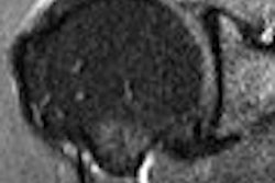
The researchers sought to compare the accuracy rate of unenhanced sonography, contrast-enhanced sonography, and MRI in displaying the anatomic features of cystic pancreatic masses greater than 1.5 mm in diameter. They retrospectively reviewed unenhanced and contrast-enhanced sonographic and MRI examinations of 33 patients who underwent resection of a cystic pancreatic mass between January 2004 and January 2006 (AJR, December 2007, Vol. 189:6, pp. 1435-1442).
Ultrasound was performed using a Sequoia 512 scanner (Siemens Medical Solutions, Malvern, PA), with contrast-specific sonographic imaging modes and low acoustic pressure (2-4 MHz coherent contrast imaging or cadence contrast pulse sequencing). For contrast studies, a 2.4-mL bolus SonoVue, (Bracco, Milan, Italy) was injected intravenously and immediately followed by a 5-mL bolus of saline solution. MRI scans were performed on a Magnetom Symphony (Siemens) 1.5-tesla scanner with a phased-array surface coil.
All unenhanced sonographic, contrast-enhanced sonographic, and MRI examinations had been performed within one week before surgery. Blinded to the final diagnosis, two radiologists reviewed the images, assessing the presence of intralesional mural nodules and septa. The researchers then calculated the sensitivity, specificity, positive predictive value, negative predictive value, and accuracy based on the correlation with surgical findings.
Contrast-enhanced sonography correctly depicted intralesional septa in 14 of 15 lesions and nodules in six of eight lesions. MRI also correctly depicted intralesional septa in 14 of 15 lesions. For nodules, MRI correctly depicted seven of eight lesions. The results for sensitivity, specificity, positive predictive value (PPV), negative predictive value (NPV), and accuracy for both modalities are shown below.
|
|||||||||||||||||||||||||||||||||||
While the difference in diagnostic accuracy of contrast-enhanced sonography and MRI was not significant (p = 0.05) in identifying septa and nodules, the researchers found that the correlation between contrast-enhanced sonographic findings and pathologic results (Rs = 0.93, p < 0.001) was significantly better than that between sonographic and pathologic results (Rs = 0.52, p < 0.0001). Interobserver agreement had a K value of 0.86-0.94.
"Our findings, however, prove that in patients in whom the cystic pancreatic mass is visible on transabdominal sonography, the results of contrast-enhanced sonography and those of MRI in detecting intralesional septa and nodules are very similar," the researchers wrote.
CEUS can be considered a complementary study for characterizing cystic pancreatic masses seen on transabdominal sonography, and can be included in the follow-up of borderline lesions, they concluded.
"In this subgroup of patients in whom a cystic mass can be visualized, contrast-enhanced sonography may be a less expensive, radiation-free, and effective imaging technique of lesion follow-up," the authors wrote.
By Erik L. Ridley
AuntMinnie.com staff writer
November 23, 2007
Related Reading
Contrast media add valuable information in US applications, May 4, 2007
DTHI boosts US performance in hepatic imaging, March 16, 2007
CEUS stars in differentiating PVT in cirrhotic patients, December 7, 2006
CEUS shines in characterizing hypoechoic focal lesions in fatty livers, November 28, 2006
CEUS aids differentiation of small liver lesions, April 3, 2006
Copyright © 2007 AuntMinnie.com