Researchers at the University of Texas M. D. Anderson Cancer Center in Houston have successfully detected heterogeneous early tumor response to head and neck radiotherapy through dynamic contrast-enhanced (DCE) MRI and diffusion-weighted (DW) MRI.
The prospective pilot study's findings also suggest that DCE-MRI and DW-MRI potentially could guide dose escalation during treatment of refractory high-risk disease.
In the study, researchers used a CT-on-rails imaging system to deliver volumetric image-guided adaptive radiotherapy to oropharyngeal cancer patients on a daily basis. All MR images were acquired using a 3-tesla MRI scanner (Excite HD, GE Healthcare, Chalfont St. Giles, U.K.) and eight-channel high-definition neurovascular phased-array coil.
In addition, CT images were used to compute dose distribution, while image registration was utilized to correlate radiation doses administered to patients on a pixel-by-pixel basis at the three-week mark, midway through their treatment, and at post-treatment via DCE-MRI and DW-MRI images.
Slow changes
"Unfortunately, structural changes in head and neck volume change relatively slowly," said Dr. David Schwartz, study co-author from M. D. Anderson Cancer Center. "Through biological changes, as a result of radiation treatment, tumor changes are emblematic of radiation response. We used DCE-MRI and vascular data to potentially quantify localized changes prior to structural changes."
Schwartz's comments came during his presentation of the pilot study at last week's annual meeting of the American Society for Therapeutic Radiology and Oncology (ASTRO) in Boston.
Researchers also obtained contiguous 5-mm thickness T1-weighted and fat-suppressed T2-weighted MR images in the axial plane as part of the data collection. They also performed a parallel imaging calibration scan, followed by a fat-suppressed spin-echo planar DW-MRI acquisition from 3-mm sections.
DCE-MRI data were acquired from identical regions every six seconds before, during, and after bolus injection of 0.1 mmol/kg gadopentetate dimeglumine delivered at 3 mL/sec.
Schwartz presented pilot study MRI data from four patients, observing and analyzing dose response relationships across all cases, which were detected more sensitively through deformable mapping versus rigid mapping of dose distributions.
K-trans calculations
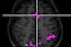
Researchers used DW-MRI and K-trans maps (the difference between baseline scans and repeat MRI scans) to identify heterogeneous early tumor response in diffusion restriction and K-trans maps within gross tumors at the three-week point of treatment.
"Image registration was critical to this project," Schwartz said. "We wished to do pixel-to-pixel spatial correlations in order to do serial K-trans mapping within patients across time, as well as do spatial correlated dose with K-trans values."
Because there is no gold standard to compare with DCE-MRI, researchers sought to create what Schwartz described as "a usable, reproducible summary of statistics that potentially could be used in a clinical trial setting ... so we settled on the mean changes of K-trans."
Researchers also relied exclusively on anatomical imaging, rather than functional imaging, "for as precise registration as possible throughout the entire course of the study," Schwartz added.
Disease response
The study noted that primary and nodule disease response patterns among the four patients were "distinct." In one representative case, 20% and 60% mean reductions in K-trans maps were observed in gross nodal disease at 34.5 Gy at three weeks and at six weeks after completing 69.2-Gy treatment, respectively.
Conversely, the study noted that K-trans maps increased within a tonsil primary at three weeks (36 Gy), followed by a post-treatment reduction in normal tissue values. MRI also identified occult gross tumors, which prompted new adaptive treatment plans.
"Even in a small pilot patient population, we were able to turn up early and late response groups" in regard to head and neck radiotherapy, Schwartz said, "but we don't quite yet know the biological and clinical significance of the earlier response according to the changes in K-trans value."
Because there currently is no published data for DCE-MRI for head and neck radiation response, Schwartz expects interest in this application to grow among radiation oncologists.
By Wayne Forrest
AuntMinnie.com staff writer
September 30, 2008
Related Reading
Breast MRI can guide radiation therapy to lymph nodes, September 22, 2008
Interest surges in use of imaging for tracking radiation therapy, July 31, 2007
Dynamic contrast-enhanced MRI shows promise in mesothelioma, September 15, 2006
Farewell to fusion: Contrast MRI edges CT in postbrachytherapy exams, May 19, 2004
Copyright © 2008 AuntMinnie.com