With the help of 3-tesla MRI, researchers in Honolulu have found indications that prenatal methamphetamine exposure accelerates brain development in an abnormal pattern, according to a study published this week in Neurology.
Such abnormal brain development, the authors wrote, "may account for the slower maturation of behavioral measures observed in neonates with prenatal methamphetamine exposure." The study, co-authored by Dr. Linda Chang with the John A. Burns School of Medicine at the University of Hawaii at Manoa, is published in the April 15 online issue of Neurology.
The researchers analyzed a sample of 29 children who were exposed to methamphetamine and 37 unexposed children between the ages of 3 and 4 years. The children were recruited from local hospitals, drug rehabilitation centers, and the local community.
Cumulative methamphetamine exposure was determined through detailed interviews of the mothers regarding the quantity of methamphetamine used on a daily and weekly basis, the duration of use, and their patterns of lifetime use. To evaluate possible environmental influences on brain development, the children's primary care providers (biologic mothers and, in some cases, legal guardians) were evaluated for their estimated verbal intelligence, mood, and socioeconomic status.
The children also were given physical and neurologic evaluations and screened for contraindications for the MRI studies.
Study criteria
Methamphetamine-exposed children included in the study were between 36 and 59 months old and had been subjected to their mothers' methamphetamine use during pregnancy or had a positive toxicology at birth.
As is standard medical practice, the young children were not sedated during their scans on a 3-tesla MRI system (Magnetom Trio, Siemens Healthcare, Malvern, PA), using an eight-channel phased-array radiofrequency coil and a diffusion-tensor imaging (DTI) technique.
The researchers scanned the children during a nap, after a meal, or while watching a favorite movie. It took as many as five sessions to image many children to acquire an adequate scan. Scans generally took 40 to 45 minutes to complete.
Methamphetamine exposure
Children with prenatal methamphetamine exposure were exposed to methamphetamine for an average of 2.5 trimesters. Of the 17 children who had records of urine or meconium toxicology, 65% of the youngsters tested positive for methamphetamine at birth.
During pregnancy, meth usage of mothers in the study averaged 58 grams, though the degree of usage varied greatly among the subjects. Although none of the mothers met criteria for any other drug dependence except for methamphetamine or nicotine while pregnant, the mothers who used meth smoked more cigarettes, drank more alcohol, and showed a trend for more marijuana use.
The 3-tesla MRI scans showed that children with prenatal exposure to methamphetamine had differences in the white matter structure and maturation of their brains when compared to children with no exposure. They also had as much as 4% lower diffusion of molecules in the white matter of their brains, specifically in the right frontal (-2.1%), right parietal (-3.9%), and left parietal white matter (-3.3%), as well as a trend toward lower diffusion in the left frontal white matter (-2.0%).
Abnormal development
Chang noted that the study's findings suggest prenatal methamphetamine exposure accelerates brain development in an abnormal pattern, and such abnormal brain development may explain why some children with prenatal methamphetamine exposure reach developmental milestones later than others.
Previous research has shown that prenatal methamphetamine exposure can lead to increased stress and lethargy and poorer quality of movement for infants.
While the Hawaiian study could not conclude definitively how prenatal methamphetamine exposure may lead to lower brain diffusion, less diffusion of molecules in white matter typically reflects more compact axonal fibers in the brain, Chang said. That occurrence is consistent with researchers' prior findings of smaller subcortical structures in children with prenatal methamphetamine exposure.
 |
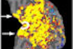
| The clinical images are representative region-of-interest placements on fractional anisotropy maps for the seven regions studied. Thalamus (E), globus pallidus (F), putamen (G), caudate (H), and white matter regions [frontal white matter (C) and parietal white matter (D)] were drawn bilaterally, while corpus callosum was drawn as indicated in the splenium (A) and genu (B). Images courtesy of Neurology and the University of Hawaii at Manoa. |
"Taken together, our findings suggest prenatal methamphetamine exposure accelerates brain development in an abnormal pattern. Such abnormal brain development may account for the slower maturation of behavioral measures observed in neonates with prenatal methamphetamine exposure," the study noted.
Follow-up research
Chang said they plan to follow the youngsters on an annual basis for several years to look for patterns of behavior and development that may be associated with prenatal methamphetamine exposure or to see if they will normalize as they become older.
The authors noted that the study's findings are confounded by the lack of drug histories on some subjects, which "may minimize or exacerbate the effects of methamphetamine."
"In addition," they wrote, "genetic and environmental influences cannot be ruled out as a source for the lower [white matter] diffusion, since the children whose mothers with greater lifetime substance use disorder probability had lower diffusivity, while estimated amount of prenatal drug exposure did not appear to influence brain diffusivity."
The researchers tried to minimize the effect of any variables that may skew the results by narrowing the age range to minimize variability on DTI measures associated with age, and by recruiting children at a young age to minimize environmental influences.
The study was funded by the National Institute on Drug Abuse and supported by the National Center for Research Resources, the National Institute of Neurological Disorders and Stroke, and the Office of National Drug Control Policy.
By Wayne Forrest
AuntMinnie.com staff writer
April 15, 2009
Related Reading
MRS detects positive neuronal changes in ex-meth abusers April 6, 2005
Crank call: Imaging exposes major brain alterations in meth abusers, February 28, 2005
MRS shows cost-benefit strength for brain tumor diagnosis, January 28, 2005
Ex-meth abusers show some reversal in brain damage, March 15, 2004
Copyright © 2009 AuntMinnie.com