There is nothing subtle about a teenage boy -- in all his unchecked and youthful glory -- hurtling down the highway in a car "borrowed" from dad. So it's not surprising that of the 160,000 thoracolumbar spine fractures that occur annually, most happen in young males involved in car collisions. But the spectrum of imaging findings in these fractures may be difficult to detect, leading a group in Maryland to catalog the most common ones in the American Journal of Roentgenology.
"Flexion-distraction, or Chance-type, fractures are often subtle on both radiography and CT, and are unstable injuries that usually do not present with a neurologic deficit," wrote the group from the University of Maryland School of Medicine and the Maryland Shock Trauma Center, both in Baltimore.
"Spine injuries are common in major blunt trauma and may be missed, or diagnosis may be delayed in polytrauma patients," added lead author Dr. Mark Bernstein, who is now at New York University Medical Center in New York City (AJR, October 2006, Vol. 187:4, pp. 859-868).
For this retrospective review, Bernstein's group looked at 53 patients with 55 separate Chance fractures of the thoracolumbar spine over an eight-year period. Most of the patients (72%) were male and sustained their injuries in car crashes (72%). At presentation, 88% had a normal Glasgow Coma Scale score of 15 with a mean injury severity score (ISS) of 19. Spinal fusion treatment was the dominant treatment protocol (60%).
All patients had anteroposterior (AP) and lateral radiographs of the thoracolumbar spine with 78% of the injuries occurring at the thoracolumbar junction, between T12 and L2. A transverse fracture through the pedicles was seen in 66% of the cases with increased intercostal spacing observed in 50%.
 |
| Chance fracture in 28-year-old woman. Cross-table lateral radiograph of lumbar spine shows fanning of spinous processes (double-headed arrow) and fracture extending through pedicle (between arrowheads) and into L2 vertebral body (single-headed arrow). |
At CT, transaxial images revealed uncovering of the articular facets secondary to the vertical distraction of the posterior elements, the authors stated. In addition, 76% of the patients had the "dissolving pedicle" sign on CT in which there is a gradual loss of definition of the pedicles.
"A close examination of the fracture patterns of CT unexpectedly reveal that a burst-type fracture with buckling or retropulsion of the posterior cortex was a common finding, seen in 48% of the patients in our series," the group wrote.
 |
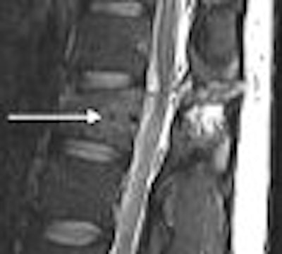
| Inversion-recovery sagittal MR images of lumbar spine in 31-year-old man. Chance fracture of L1 shows marked high signal in interspinous ligaments and soft tissues (arrowheads). Bone marrow edema (arrows) is seen in pedicle and vertebral body. |
MR showed marked soft-tissue damage through the posterior elements and surrounding soft tissues in all patients. "MRI evaluation provides information regarding the integrity of the posterior and middle-column ligaments, annulus fibrosis, and spinal cord in neurologically injured patients," they added.
Associated with the Chance-type fractures were intra-abdominal injuries, seen in 40% of their patients, the authors wrote. This connection between abdominal injuries and thoracolumbar fractures first became apparent in the 1960s when seatbelts became standard in vehicles, the authors explained.
 |
| Inversion-recovery sagittal MR images of lumbar spine in 31-year-old man. Low-signal fracture line seen centrally in posterior vertebral body (arrow) with surrounding edema represents MRI sandwich sign. Bernstein MP, Mirvis SE, Shanmuganathan K. "Chance-Type Fractures of the Thoracolumbar Spine: Imaging Analysis in 53 Patients" (AJR2006; 187:859-868). |
"We hypothesize that the fulcrum, or axis of rotation, begins at the site of the lap belt … pressed against the anterior abdominal wall at the time of impact and sudden deceleration," they wrote.
As for why the thoracolumbar spine may be particularly prone to injury, especially during car crashes, the authors pointed to biomechanics, including the transition from the relatively rigid thoracic spine to the more mobile lumbar spine, and the loss of stability in the thoracic spine because of articulation to the rib cage and sternum.
A scientific paper at the 2006 RSNA meeting in Chicago will offer more on the biomechanics of the spine, defining the normal angulation at the thoracolumbar junction (SST17-03). Also, a pictorial review will highlight musculoskeletal complications after traumatic spinal cord injury (education exhibit LL-MK5249).
By Shalmali PalAuntMinnie.com staff writer
November 9, 2006
Related Reading
Pilot study spots 2 factors that signify acute intrathoracic injury after blunt trauma, May 22, 2006
CT more sensitive than spinal fracture x-ray, but has limits, January 30, 2006
Copyright © 2006 AuntMinnie.com