An abnormal acromial undersurface can be the cause of rotator cuff irritation that leads to subacromial outlet impingement. But getting an accurate view of the area hasn't been a straightforward exercise -- at least not in the medical literature.
Now researchers from Austria are attempting to resolve this historical challenge with a new comparison of x-ray and MR imaging of the acromion.
The findings and recommendations from Dr. Marius Mayerhoefer and colleagues from the Medical University of Vienna and the Orthopedic Hospital Vienna-Speising are published in the American Journal of Roentgenology (February 2005, Vol. 184:2, pp. 671-675).
The role of acromial shape in shoulder problems was first identified in the mid-1980s by Dr. Louis Bigliani, now chairman of orthopedic surgery at Columbia University in New York City.
Based on cadaver specimens, Bigliani described three different types of acromion: flat type 1, curved type 2, and hooked type 3. Both curved and hooked acromions are regarded as abnormal variants, with the latter particularly implicated in the development of outlet impingement.
Outlet-view radiographs became the gold standard for assessing acromial shape because Bigliani and his co-authors initially found a good correlation between those images and rotator cuff tears, the AJR authors noted.
 |
| Mathematic determination of acromial shape. Line connecting most caudal points of acromial undersurface is drawn on parasagittal T2-weighted MR image, and with help of two orthogonal lines, acromion is divided into three segments of equal length. |
"Subsequent studies, however, have shown that correlation decreases if the radiographs are obtained by different technologists and that minor changes of the angle of the central beam may influence considerably the depiction of acromial shape by these radiographs," they wrote.
While MRI doesn't have such a projection-error problem, "the appearance of the acromial shape is highly dependent on the position of the MRI plane and therefore may change from slice to slice," the authors wrote.
Thus, the researchers also set out to perform the first published comparison of the various slice positions recommended in the literature.
 |
| Mathematic determination of acromial shape. Then on parasagittal T2-weighted MR image, angle between anterior third and posterior two-thirds is measured. In this case, the anterior angle of deflection is 180° - 168° = 12°, indicating slightly curved (type 2) acromion in S-2 position. |
Their study looked retrospectively at 61 patients with clinically confirmed impingement syndrome who underwent arthroscopic acromioplasty after conservative treatment had failed.
All of the patients were examined with outlet-view radiographs, taken by various technologists, prior to acromioplasty. The acromial shape revealed by those radiographs was ascertained by consensus of one radiologist and an orthopedic surgeon, and then compared with the shape seen during surgery.
The patients also underwent MRI prior to surgery. Parasagittal T2-weighted MR images, perpendicular to the supraspinatus tendon, were obtained on a 1-tesla MRI scanner with a dedicated shoulder surface coil, using a TR of 2,000 msec, a TE of 140 msec, a 256 × 256 matrix, and a slice thickness of 4 mm.
The researchers constructed 3D models of the acromions from the MR data. Acromial shape was again determined by consensus and compared with surgical findings.
   |
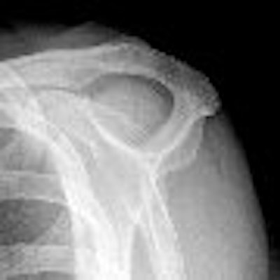
| A 63-year-old man with impingement syndrome of shoulder. Whereas outlet view radiograph (top) depicts curved acromion (type 2), MR image (middle) in position S-1 reveals blunt acromial hook (type 3), which is also confirmed by 3D model (above) and intraoperatively. Mayerhoefer ME, Breitenseher MJ, Roposch A, Treitl C, Wurnig C, "Comparison of MRI and Conventional Radiography for Assessment of Acromial Shape" (AJR 2005; 184:671-675). |
"To provide a reliable reference for evaluation of the different MRI slices and outlet-view radiographs, we used only those 56 acromions (90.3%) with agreement on acromial shape between intraoperative findings and the 3D model," the authors wrote.
From those 56 datasets, the researchers then extracted three MRI slices equivalent to those favored in various earlier studies.
One slice, dubbed S-1, was located 4 mm from the lateral margin of the acromion; another was located just lateral to the acromioclavicular joint (S-2); the third was just medial to S-2, depicting the most lateral portion of the acromioclavicular joint (S-3).
The researchers then calculated the sensitivity and specificity of those three slices and for the outlet-view radiographs.
"For determination of acromial shape, outlet-view radiographs are superior to any single MRI slice position," the authors concluded.
However, "the best results were achieved through a combination of measurements in slice positions S-1 and S-2 (calculation of arithmetic mean of angle measurements). With a kappa coefficient of 0.66, this two-slice combination appears to be superior to outlet-view radiographs and may, therefore, be recommended, if time permits."
By Tracie L. ThompsonAuntMinnie.com staff writer
February 21, 2005
Related Reading
Standard and extremity MR bear brunt of shoulder imaging, November 17, 2004
Shoulders bear brunt of water polo's demands, August 20, 2004
Ultrasound of muscles adds vital information on rotator cuff condition, August 16, 2004
Getting the most from shoulder positioning, December 24, 2002
Dynamic MRI techniques detail patterns, problems in shoulder motion, June 28, 2000
Copyright © 2005 AuntMinnie.com