Researchers from Wake Forest University Baptist Medical Center in Winston-Salem, NC, are developing a method of cancer treatment that uses lasers and MRI to illuminate tiny nanoparticles and destroy tumors with the ensuing heat.
Co-developer Xuanfeng Ding, from Wake Forest's department of physics, presented details of the project on Wednesday at the American Association of Physicists in Medicine (AAPM) annual meeting in Philadelphia.
The technique uses iron-containing multiwalled carbon nanotubes (MWCNTs), which are threads of hollow carbon that are 10,000 times thinner than a human hair. The work utilizes an experimental technique for treating cancer called laser-induced thermal therapy (LITT), which uses energy from lasers to heat and destroy tumors.
LITT works by way of certain nanoparticles, such as MWCNTs, that can absorb the energy of a laser and convert it into heat. If the nanoparticles are activated while they are within a tumor, they will produce energy as heat and kill the cancerous cells.
According to the researchers, the problem with LITT is that while a tumor may be clearly visible in a medical scan, the particles are not. They cannot be tracked once injected, which could place a patient in danger if the nanoparticles were activated away from the tumor, because the aberrant heating could destroy healthy tissue.
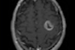
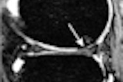
The Wake Forest researchers, however, have shown that it is possible to visualize the particles in the MRI scanner to allow imaging and heating at the same time. The key is the MWCNT particles with iron, which become visible on MRI.
Using tissue containing mouse tumors, the researchers were able to view the nanoparticles with MRI, track as they approached a tumor, and target them with a laser, destroying the tumor in the process.
Related Reading
fMRI can help predict tumor response, July 12, 2010
Study offers strategy for managing incidental findings on fMRI, July 6, 2010
Copyright © 2010 AuntMinnie.com