MRI is more sensitive in identifying breast cancer than mammography, but its role in screening has been controversial and its diagnostic use varied. Better guidelines are needed to increase the modality's value in breast imaging, according to a presenter at the 2006 American Society of Breast Disease (ASBD) symposium in Las Vegas.
MRI particularly shines when mammography is inconclusive, such as in women with dense breasts, small lesions, or lobular carcinoma, said Dr. Steven Harms, a radiologist at Breast Center of Northwest Arkansas in Fayetteville, AK.
Advances in MRI technology have made tumor characterization even clearer. Harms discussed the advantages of rotating delivery of excitation off-resonance (RODEO) MRI while Dr. Alan Hollingsworth from Iowa talked about MRI screening intervals.
MRI advances
Interpretation of what can be a "mammographer's nightmare" is much easier using contrast agents in conjunction with high-resolution MRI systems like RODEO (Aurora Imaging Technology, North Andover, MA), said Harms, who serves as the medical director for Aurora. The technology is a proprietary pulse sequence that provides fat suppression and magnetization transfer contrast for a high-resolution image.
"Even if you think you got a really good mammogram, sometimes MRI helps in clarifying things," Harms said.
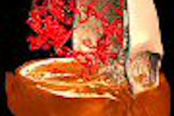
Precontrast, dense breast images may be riddled with bright spots from cysts and therefore very difficult to interpret. But once the contrast agent washes in, ductal tissue is suppressed by the magnetization transfer contrast and enhancing tumors are relatively easy to spot.
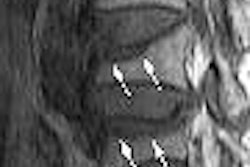
Lobular carcinoma, which accounts for 10% of breast cancers and is bilateral in up to 30%, is difficult to detect on physical examination or mammography. As a result, lobular carcinoma is often underdiagnosed and overtreated, Harms said, which gives MRI the advantage for defining the disease.
While low-resolution MRI can miss this diagnosis because of volume averaging, RODEO clearly depicts the extent of the lobular carcinoma allowing lumpectomy rather than mastectomy, he said.
MRI also offers advantages for ductal carcinoma in situ (DCIS), Harms added. The extent of DCIS is difficult to assess on physical exam or mammogram, and it is routinely missed by low-resolution dynamic MRI studies because volume averaging limits visualization.
Harms cited recent trials indicating that MRI picks up four times as much DCIS as mammography (Breast Journal, November-December 2005, Vol. 11:6, pp. 379-390; Breast Journal, September-October 2004, Vol. 10: sup. 2, pp. S1-S16).
Finally, Harms touched on spiral MRI, calling it an emerging technology that "is going to tremendously help in finding those small lesions." Spiral MRI makes a 1.5-tesla magnet the equivalent of a 4.5-tesla magnet, giving it three times the signal-to-noise ratio in one-third the time by more efficiently filling voxels, he explained. "It’s as good as it gets in MR," he said.
Screening interval recommendations
Several groups, including the American Cancer Society, recommend that high-risk women (genetic mutations carriers, family history) consider ultrasound and MRI, in combination or along with screening mammography, but there are no recommendations for the interval at which MRI testing should occur.
The best strategy may be an individually tailored interval of MRI combined with annual mammography, said Dr. Alan Hollingsworth, medical director at Mercy Women's Center in Cedar Rapids, IA.
"One can posit that capturing the other half through improved screening would double the current mortality reduction imparted by mammography alone," Hollingsworth said.
He described the protocol at his institution based on a risk and breast density stratification. An average-risk patient gets 1 point, while elevated or high-risk patients get 2 points and very high-risk patients get 3 points. These risk levels are based on his group's published management strategy (American Journal of Surgery, March 2004, Vol. 187:3, pp. 349-362).
Breast density is classified according to American College of Radiology Breast Imaging Reporting and Data System (BI-RADS) guidelines. Predominantly fatty breasts get 0 points. Scattered fibroglandular densities get 1 point. Heterogeneously dense breasts get 2 points, and extremely dense breasts get 3 points.
These density and risk points are combined for each individual's recommended MRI screening interval score. A patient with 2 points should have MRI screening every three years while a patient with 3 to 4 points should have it every two years. Women with 5 to 6 points should receive yearly MRI screening in addition to yearly mammography.
If a patient has only 1 point total, no MRI screening is recommended. If a patient has level 3 or 4 breast density, Hollingsworth also advised ultrasound screening in the "off years" between MRI screenings. "The bulk of patients interested in MRI screening fall in the biennial range," he said.
Although MRI screening is tremendously cost-ineffective, this should come as no surprise since mammographic screening itself is not cost-effective, Hollingsworth said.
He referred clinicians to the International Working Group on Breast MRI for more information on implementing a MRI screening program (Breast Journal, September-October 2004, Vol. 10: supplement 2, pp. S1-S2).
By Crystal Phend
AuntMinnie.com contributing writer
May 26, 2006
Related Reading
Aurora installs breast MRI at Texas center, April 5, 2006
Aurora adds Dallas install, March 14, 2006
MR mammography: Screening tool or diagnostic adjunct? February 9, 2006
Larger lesions on breast MRI equal greater chance of cancer, February 8, 2006
Adding MRS to MRI ups specificity for finding breast lesions, December 16, 2005
Copyright © 2006 AuntMinnie.com