PHOENIX - Three-tesla MRI offers improved signal-to-noise and contrast-to-noise ratios compared with 1.5-tesla for body imaging, according to a presentation at the 2006 Society of Computed Body Tomography and Magnetic Resonance (SCBT/MR) meeting. However, several issues such as cost, weight, safety, and modified imaging protocols may mitigate these advantages.
"It seems like body imagers have really found their niche at 1.5 tesla," said Dr. David Bluemke, who is from the department of radiology at Johns Hopkins Hospital in Baltimore. "It provides an excellent combination of speed (and) spatial and contrast resolution. The image quality is very nice, with MR angiographic and multiplanar evaluations being readily available."
Bluemke, an associate professor of radiology and medicine and clinical director of MRI at Johns Hopkins, discussed the advantages and disadvantages of 3-tesla imaging of the abdomen and pelvis, compared with 1.5-tesla, during the SCBT/MR meeting on Monday.
The challenges of body MRI, according to Bluemke, are that the signal-to-noise ratio (SNR) is often low and that high spatial and temporal resolution are needed, requiring breath-hold imaging. The trade-offs are that a smaller pixel size provides low SNR and that high temporal resolution delivers low SNR or large pixel size.
Three-tesla MRI provides four times more signal than 1.5-tesla MRI; however, the noise also increases twofold. The net gain, Bluemke said, is that twice as much signal is available at 3 tesla compared with 1.5 tesla. As appealing as this may sound, the hardware seems to have outstripped software capabilities in regard to body imaging, Bluemke noted.
"There are very few applications that can exploit the benefits (of the increased SNR) of 3-tesla body imaging, but they are being developed," he said.
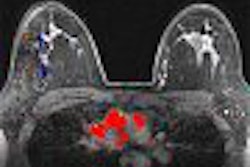
The contrast-to-noise ratio (CNR) is also improved at 3 tesla compared with 1.5 tesla. At 3 tesla, the T1 relaxation time is longer. The longer T1 of background tissues improves target-to-background contrast and provides for a potential decrease in contrast dose, Bluemke.
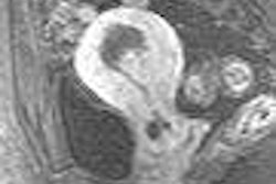
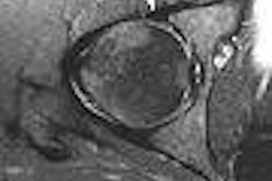
Prostate exams conducted with 3-tesla MR, both imaging and spectroscopy, show potential for greatly benefiting from the technology, he said. In a recent study in Radiology (January 2006, Vol. 238:1, pp. 184-191), researchers reported an accuracy of 94%, a sensitivity of 88%, and a specificity of 96% for prostate cancer staging using 3-tesla endorectal MRI. Researchers using 1.5-tesla equipment report accuracies of 54% to 88% conducting similar exams, according to Bluemke.
MR prostate spectroscopy conducted at 3 tesla shows better spectral resolution and better distinction of metabolites, compared with 1.5-tesla exams, Bluemke said.
"The increased SNR allows for improved detection of metabolites," he said.
But the high field strength is not without its challenges. Image distortion is a particular problem for 3-tesla and higher MR scanners. An inhomogeneous magnetic field (the B0 effect) is caused by susceptibility differences at air-tissue interfaces.
This results in both signal loss and geometric distortion of images from the modality, and this problem is more severe in the abdomen than the brain. A potential solution is to improve shimming in these devices, Bluemke said. Pulse sequences also need to undergo adjustments and optimization for 3-tesla use, which may necessitate new imaging strategies.
Safety issues are another concern for widespread deployment of 3-tesla body imaging. The magnetic fringe field of these systems is much greater than those of 1.5-tesla units. In addition, devices labeled as "MR Safe" at 1.5 tesla may not necessarily be safe at 3 tesla, Bluemke said.
Acquisition, deployment, and ongoing costs are other issues bearing scrutiny for those seeking to implement 3-tesla MR. The price of this modality is roughly double that of a comparable 1.5-tesla MR. In addition, the weight of the system is also doubled, a significant factor when installing the technology. Cryogen consumption at 3 tesla is approximately three times that of 1.5 tesla on an hourly basis, Bluemke said, which also adds to the cost of maintaining the system.
"I think most of the body imagers out there are incredibly satisfied with imaging at 1.5 tesla," he said.
Regarding 3-tesla applications, "we've got to do the investigations and determine how well these are going to work," Bluemke noted. "There are a lot of really promising areas out there."
By Jonathan S. Batchelor
AuntMinnie.com staff writer
April 4, 2006
Related Reading
Post-UAE MRI demonstrates symptom control for adenomyosis, March 15, 2006
Algorithms for staging prostate cancer are improved with MRI, February 28, 2006
High resolution, fast acquisition gives 3D MRA edge in abdomen, January 20, 2006
Low-field MR with the right coil tops 3-tesla MR for prostate cancer imaging, December 15, 2005
The 3-tesla MRI swap: Why it's not a simple upgrade, January 27, 2005
Copyright © 2006 AuntMinnie.com