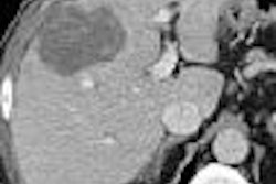
NEW ORLEANS - The number of liver lesions detected by MDCT presents a challenge for patient management, especially when the patient already has a primary malignancy. But MRI can significantly reduce the number that might otherwise be headed for biopsy, according to a study presented at the American Roentgen Ray Society (ARRS) meeting.
Dr. Nagaraj Holalkere and colleagues from the Massachusetts General Hospital in Boston looked at the added value of MRI on patient management in the wake of burgeoning numbers of MDCT-detected liver lesions.
"Although we know that 80% to 90% of the focal liver lesions -- even in patients with cancer -- are benign, characterization of lesions has a significant impact on patient management," Holalkere noted.
"Routinely, when we see a lesion in a patient with cancer, we follow on repeat serial CT to see if the lesion is stable or not," he said. "This kind of approach has some limitations because there are no standard guidelines for such serial CT studies. It requires multiple studies, and it's time-consuming."
The researchers evaluated 112 patients with known primary and suspected liver lesions who underwent liver MRI within six weeks of an MDCT exam.
The MDCT exams were performed on four- or 16-slice scanners with nonionic contrast in the portovenous and equilibrium phases. Only selected patients were scanned in the arterial phase, based on their primary cancer.
MR imaging was performed on a 1.5-tesla scanner with gadolinium-DPTA enhancement in all three phases.
The radiologists recorded their diagnostic confidence for both the MDCT and MR interpretations on a scale of 1 to 5, with 1 as "definitely benign" and 5 as "definitely malignant."
Holalkere presented a chart showing that readers were much more confident when looking at the MR images. "You can see the patients were mainly categorized by MDCT to either 'indeterminate' or 'probably malignant,' whereas MRI could confidently characterize most of them as either 'definitely benign' or 'definitely malignant' with very few indeterminate lesions," he said.
As confirmed by histopathology or follow-up imaging of at least six months, 51% of the patients had lesions that were malignant. But many more of them would have undergone an invasive procedure based on the CT findings alone.
The CT findings suggested 83% of the patients needed biopsies -- a call justified in only 61% of the cases. By comparison, MR recommended biopsy for just 55% of the patients, which would have been justified in 87% of their cases.
Liver MR has significantly higher accuracy in characterizing liver lesions detected on MDCT in patients with known malignancy, and can also have a significant impact on the management of those patients, the researchers concluded.
"By performing an MRI we could reduce the invasive procedures by almost 30% as compared to MDCT," Holalkere said.
By Tracie L. Thompson
AuntMinnie.com staff writer
May 19, 2005
Related Reading
Ultrasound contrast enhances liver imaging, May 11, 2005
Contrast US pinpoints focal liver lesions, results in cost-savings, March 25, 2005
Breath-hold technique comparable to respiratory-triggered MR for liver imaging, March 16, 2005
Exam technique critical in liver ultrasound, October 22, 2004
Copyright © 2005 AuntMinnie.com