Routine bone scans may be safely eliminated and replaced with FDG-PET/CT in the evaluation of higher-risk children, adolescents, and young adults with Hodgkin's lymphoma.
The conclusion comes from new research from St. Jude Children's Research Hospital in Memphis, TN. Dr. Barry Shulkin, co-author of the study and director of the division of nuclear medicine at St. Jude, presented results at the 2008 annual meeting of SNM in New Orleans.
The purpose of the research was to evaluate the roles of FDG-PET/CT and skeletal scintigraphy in the management of children, adolescents, and young adult patients with Hodgkin's lymphoma.
The study included 18 patients, ranging in age from seven to 24 years. Researchers looked for patients who had received both an FDG-PET/CT and bone scan within two weeks of each other. Among those patients, the study found 38 scan pairs -- 11 patients (11 scans) were studied at diagnosis, while seven patients (27 scans) were studied with known Hodgkin's lymphoma or were suspected to have relapsed.
Two physicians independently reviewed the bone and FDG-PET/CT scans, with a third physician resolving any discrepancies.
Researchers found six patients, with a total of nine scans, that were positive on both FDG-PET/CT and the conventional bone scans. Both the FDG-PET/CT and bone scans concurred on a negative finding for nine patients.
 |
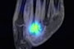
| Images of a 14-year-old boy with nodular sclerosing Hodgkin's lymphoma at presentation. The left panel shows a posterior projection image from an FDG-PET/CT scan with many areas of abnormal osseous and nonosseous uptake, in particular L2, right iliac crest, and left inferior acetabulum. The top right panel is a bone scan posterior planar view of the chest and upper abdomen, which shows only equivocal, minimally elevated uptake in the left aspect of L2. The bottom panel is a transverse fusion PET/CT image, which localizes the abnormal uptake in L2 to the left posterior elements. Images courtesy of St. Jude Children's Research Hospital and Dr. Barry Shulkin. |
 |
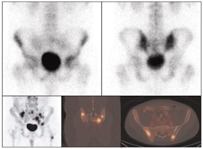
| Images of a 23-year-old man with relapsed nodular sclerosing Hodgkin's lymphoma; FDG-PET shows a greater extent of disease involvement. The top images show anterior (left) and posterior (right) spot views of bone scan, which indicate mildly increased uptake in the left acetabulum and right inferior pubic ramus. The bottom panel illustrates FDG-PET posterior maximal intensity projection (left), coronal fusion (center), and transverse fusion (right) images, with many focal areas of abnormal uptake in the pelvis and proximal femurs. |
Shulkin acknowledged some limitations of the study, noting that FDG-PET is "inherently three-dimensional. We had far higher data collection, it is attenuation corrected, and it can identify both cortical and medullary abnormalities. The bone scan, on the other hand, was done with a conventional gamma camera; it was planar; and could identify only cortical abnormalities."
He added that a better "state-of-the-art" comparison would be FDG-PET/CT and sodium chloride PET/CT and/or bone SPECT/CT.
In conclusion, the study found that all abnormal foci of uptake seen on bone scintigraphy representing metastatic disease also were evident through FDG-PET/CT scanning.
"We found that additional osseous foci of abnormal uptake were evident on FDG-PET/CT compared to the bone scan," Shulkin added, "and we conclude that in higher-risk patients routine bone scans can be safely eliminated in the evaluation of patients with Hodgkin's lymphoma."
By Wayne Forrest
AuntMinnie.com staff writer
June 30, 2008
Related Reading
SPECT/CT offers on-target diagnosis in noncancerous bone disease, March 23, 2007
Scintigraphy pegs potential responders to plantar fasciitis treatment, October 25, 2006
Experimental CAD scheme makes quick work of bone scintigraphy analysis, June 5, 2006
Bone SPECT identifies patients who would benefit from facet joint injection, February 15, 2006
Copyright © 2008 AuntMinnie.com