Plantar fasciitis is the most common cause of heel pain but the differential diagnosis can include Achilles tendinitis, retrocalcaneal bursitis, and calcaneum fracture. Bone scintigraphy is often used to identify the source of heel pain, and the modality may play a predictive role in treatment as well, according to a study in the Journal of Nuclear Medicine.
"Injections of corticosteroids or local anesthetics remain a convenient form of therapy," wrote Dr. Clayton Frater and colleagues. "Bone scintigraphy has also been shown to be an accurate guide in selecting injection sites.... We present scintigraphic criteria that can help identify potential responders (to therapy)..." (JNM, October 2006, Vol. 47:10, pp. 1577-1580).
Frater is from the University of Sydney in Australia. Other co-authors are from the University of New South Wales and Concord Hospital, both in Sydney, as well as King's College in London.
For this study, scintigraphy scans from 24 patients diagnosed with plantar fasciitis, and treated with local injection of steroids or anesthetics, were included. In addition, scans from 10 patients with other conditions were thrown into the mix to test the specificity of scintigraphy.
Imaging was done with a high-resolution collimator affixed to a gamma camera (Millennium or 400AC, GE Healthcare, Chalfont St. Giles, U.K.) after a 900- to 1,000-MBq injection of Tc-99m methylene diphosphonate. Blood-pool images of the plantar aspect of both feet were obtained, as were delayed images of the anterior, posterior, lateral/medial, and plantar aspects of the feet.
Patient responses were followed up at four to five weeks. Abnormalities of the blood-pool phase were characterized as focal calcaneal hyperemia, extension into the proximal third of the plantar fascia, diffuse involvement of the plantar fascia, or no evidence of hyperemia within the plantar fascia. Delayed images were classified based on the degree of calcaneal uptake.
In total, 32 injections were performed in 24 patients with complete or near relief from pain reported in 20 feet. Of the 20 that responded to treatment, 14 feet showed focal hyperemia and six showed minimal extension, the authors stated, adding that they found a significant difference between patients with focal hyperemia and diffuse blood-pool abnormalities (p = 0.004).
 |
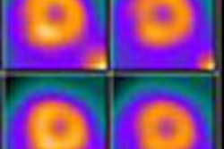
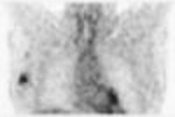
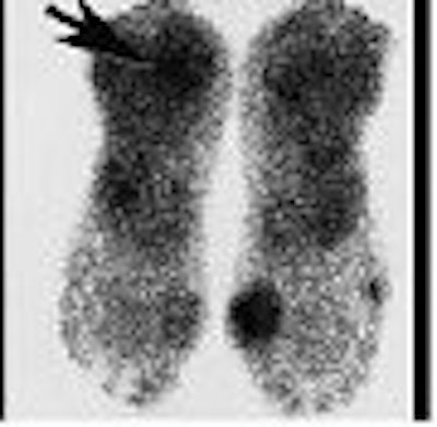
| Scan classification showing blood-pool phase at top and delayed phase at bottom: focal hyperemia in plantar fascia and marked delayed uptake in inferior calcaneum (A), hyperemia in proximal third and mild delayed uptake (B), and diffuse hyperemia and moderate delayed uptake (C). Arrowheads indicate hyperemia; arrows indicate delayed uptake. Copyright © 2006 by the Society of Nuclear Medicine. |
On the delayed images of the 20 responders, eight feet demonstrated mild inferior calcaneal uptake, six showed moderate uptake, and six had severe uptake.
"The blood-pool studies had good reproducibility, with a k-value of 0.64 (95% confidence interval [CI]). Agreement on reporting the delayed images was also good, with a k-score of 0.66 (95% CI)," the authors wrote. Interobserver agreement was fair with a k-value of 0.55, they reported.
"Focal calcaneal hyperemia is clearly associated with a high success rate for injection, with extension of hyperemia into the proximal soft tissues being associated with a response in only 50% and diffuse hyperemia with no response," the authors concluded. Using imaging criteria, clinicians can rule out patients who would not benefit from injection therapy, they added.
By Shalmali Pal
AuntMinnie.com staff writer
October 25, 2006
Related Reading
MRI, US take on diagnostic challenge of PAI in soccer players, August 18, 2006
US demonstrates continuity between lesion, plantar fascia, December 17, 2002
Copyright © 2006 AuntMinnie.com