Nuclear cardiology modalities are seeing a resurgence of late, which is not to say the most widespread heart-imaging modalities were in decline. They weren't, at least not yet. Still, recent technical improvements on multiple fronts, from detector technology to hybrid modalities like SPECT/CT and PET/CT, seem to have sharpened the modalities' future prospects as they have sharpened the images themselves.
Of course competing modalities, from which nuclear medicine has borrowed, are getting better, too. Practitioners of every modality from multidetector CT to high-field MRI and echo have sought heart imaging applications they could perform and perhaps master.
But many promising alternative technologies remain undelivered in 2006. MRI was touted as a one-stop cardiac imaging solution 23 years ago at an American College of Cardiology meeting; however, the scenario has yet to unfold in real-time, said nuclear cardiology expert Dr. Manuel Cerqueira at the recent 2006 American College of Cardiology Foundation's Integrated Imaging and Clinical Cardiovascular Practice meeting in San Francisco.
Then there is 3D stress echocardiography, "which I'm waiting for," said Cerqueira, who is a professor of radiology and the chair of molecular and functional imaging at Cleveland Clinic Lerner College of Medicine of Case Western Reserve University in Cleveland, OH.
"We need to talk about the future, but at the same time take a pragmatic focused approach ... and look at what is really required to make a technique usable," Cerqueira said.
To succeed, a heart imaging modality must be accurate and reproducible, and it must be able to answer the diagnostic question, he said. It must be cost-effective, technically feasible in all patients who need it, and deliverable.
"(If) high-end academic centers can do studies but you can't do it in a small hospital or private office, no matter how good the technique is, it's not going to work," Cerqueira said.
A new technology must answer diagnostic questions in the shortest possible time with the greatest accuracy, he said. Will SPECT and nuclear medicine be able to fulfill those requirements in the future?
All indications are that they will. SPECT works for both diagnosis and prognosis, which PET has not delivered, he said, and more evidence-based literature support its effectiveness than any other modality. SPECT has effective quality control and remains profitable despite recent reimbursement cuts.
Yet it does have limitations, including breast shadowing in women and high attenuation in the diaphragm, Cerqueira said. Technetium-based SPECT radiotracers scatter into the liver and gut, which adds counts to the inferior wall, creates display problems with normalization, and produces ramp filter artifacts. A loss of resolution occurs as a function of distance. All of these issues can complicate interpretation, especially in women and obese patients.
 |
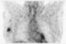
| An infant with left main artery arising from the pulmonary artery underwent surgical correction at three months. At five months the baby was crying after eating. Rest and dipyridamole stress (above) shows perfusion abnormality. At a second surgery the site where the artery was reattached was found to be kinked and corrected. At two-months follow-up, post percutaneous transluminal coronary angioplasty with resting PET (below) showed completely normal perfusion; patient is clinically stable with no ischemia, and ventricle had shrunk to normal size. PET demonstrates significantly more information density over time compared to SPECT, also providing superior scatter correction and resolution recovery. All images courtesy of All images courtesy of Dr. Manuel Cerqueira. |
 |
"We can't keep doing 6 to 8 million (nuclear cardiology) studies a year without some innovation," he said. "But I think innovation is going to result in improved accuracy, attenuation and scatter correction, and resolution recovery. The ability to measure absolute myocardial blood flow -- with PET studies, we should be able to do that."
Instrumentation is going to have one of the single biggest impacts on nuclear cardiology in the near term, he said, and its influence will come from both ends of the spectrum. On one side are smaller, faster, and less expensive camera systems that can easily be installed in a small hospital or private office. On the other end are heavy but highly capable hybrid modalities.
Dedicated office cardiac imaging systems include the Cardius XPO series gamma cameras (Digirad, Poway, CA), the CardioMD (Philips Medical Systems, Andover, MA) the Ventri (GE Healthcare, Chalfont St. Giles, U.K.) the c.cam series (Siemens Medical Solutions, Malvern, PA), the t.cam Cardio series (Toshiba America Medical Systems, Tustin, CA), and the maiCAM180 (Mid-Atlantic Imaging, Columbia, MD).
At the other end of the spectrum, hybrid PET/CT and SPECT/CT systems are large, powerful, and can be had for a million dollars or so if you get a really good deal, he said.
The small stuff
Dedicated nuclear cardiology systems can cost as little as $200,000 -- less than most echocardiography systems, Cerqueira said. And a smaller footprint is just one advantage; they also allow "very fast throughput, patients are in and out faster, and with shorter acquisition times you're going to have decreased patient motion," he said.
These systems can also be easily paired with CT scanners for attenuation correction. In fact, the broad shift to 64-slice CT scanning is making a lot of four-detector CT scans available, which are perfect for doing CT attenuation correction in combination with a small-footprint SPECT system, he said.
Brilliant but costly
One investigational technology that's gotten excellent results are SPECT cameras based on cadmium zinc telluride (CZT) crystals. CZT technology was originally developed "to detect radioactive materials and atomic bombs weeks and months after testing was done," Cerqueira noted.
Gamma camera developer Digirad of Poway, CA, explained that its predecessor firm San Diego Semiconductor (later Aurora) developed CZT-based radiation detectors in 1985, beginning full-scale production in 1991 when it received a U.S. military contract to monitor Russian warheads following the anticipated U.S. signing of the SALT III Treaty, which never occurred. Finding itself without a market in 1993, the company turned to nuclear medicine, producing the first solid-state CZT-based scanner in 1996.
But "the cost then, and I would argue yet today, is just too expensive for a commercial medical device," said Randy Weatherhead, Digirad's senior vice president of sales and marketing. In fact, CZT-based cameras are five to 10 times as costly to build as the cesium iodide thallium [CsI(Tl)] PIN diode systems that have been used in Digirad cameras since it abandoned CZT in 1998.
CZT is no more sensitive than CsI(Tl)-based systems, according to Richard Conwell, senior vice president of technology for Digirad, and leader of the team that developed the CZT scanner. "The only advantage (CZT) has is in energy resolution -- the ability to reject scatter photons," resulting in images that are "five to 10 times better in contrast" compared to other materials.
But with the end of the Cold War in the late 1980s, radiation detector vendor Aurora (now Digirad) found itself without a market before deciding get into healthcare and develop CZT-based nuclear medicine scanners.
Digirad abandoned the solid-state CZT crystals due to cost and availability concerns in favor of a CsI(Tl) PIN diode system. Two other firms are building some CZT-based scanners, including D-SPECT camera by Tirat Hecarmel, Israel-based Spectrum Dynamics and CardiArc of Lubbock, TX.
CardiArc received U.S. Food and Drug Administration (FDA) approval for its CardiArc scanner in February, but cautions on its Web site that initial production runs will likely use sodium-iodide detectors, which the company stated was similar in performance to CZT, due to the high cost and sporadic availability of CZT.
Spectrum is sticking with CZT. "To the best of my knowledge for human clinical systems, Spectrum Dynamics is the only company that's using exclusively CZT," the company's vice president of marketing, Josh Gurewitz, told AuntMinnie.com. The D-SPECT imager, which goes into beta production at the end of 2006 and will ship commercially next year, will only be offered in a CZT model, he added.
The high-density CZT crystals are sensitive to a wide range of energies, provide rapid acquisition and better resolution compared to conventional scanners, offering the potential for lower doses of radiotracers or new combinations of tracers, Cerqueira said. Scans in patients have yielded "very high sensitivity and very high count rates to acquire studies in approximately three to five minutes as opposed to the 15 to 20 minutes currently" needed with conventional systems, he noted.
Spectrum's D-SPECT scanner yielded more than 10 times the sensitivity and more than twice the spatial resolution of conventional scintillator-based gamma cameras in one study.
Ring detector systems are another useful development in some dedicated cardiac imaging systems, according to Cerqueira.
"Rather than the camera rotating, a ring goes around the patient," he explained. The D-Spect scanner has "ring detectors which oscillate and increase count rates. The aperture arc acts as collimator, which is oscillating, and the detectors allow you to get a very rapid study so you can get high counts, high sensitivity, high spatial resolution, and improvements in overall efficiency."
 |
| The CardiARC scanner has an arc-shaped detector that enables all detector pixels to be active simultaneously, providing dense angular sampling. |
Cerqueira showed CZT SPECT's capabilities in a difficult-to-image patient, a 5-ft tall 276-lb woman whose conventional SPECT results were normal. A subsequent CZT-based scan, acquired in 3.3 minutes, showed an inferior wall perfusion defect that was shown at catheterization to be a right coronary lesion.
"So there is potential for (detecting) these lesions even in challenging patients with good quality in a short time," he said. It's also cost-effective and very practical for the office setting.
 |
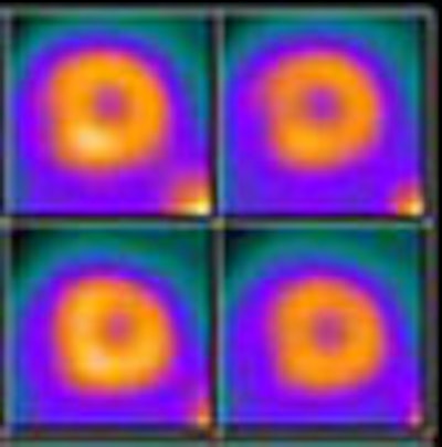
| A difficult-to-image female patient, 5 ft tall and 276 lbs, underwent conventional cardiac SPECT (top row) followed by CZT-based SPECT (CardiArc) (bottom row). The conventional study, acquired in 10.6 minutes looks fairly normal. The CZT-based SPECT, acquired in 3.3 minutes, shows an inferior wall perfusion defect that was shown to be a right coronary lesion at catheterization. |
The big guns
For facilities with money and square feet to spare, high-end hybrid systems that combine SPECT or PET with CT can overcome many of the shortcomings that limit nuclear medicine's usefulness in the most difficult-to-image cases.
But with imaging power come weight and cost. It's not just size that makes the big systems expensive -- perhaps $1 million if you get a good deal, Cerqueira said. Siting them can add significantly to the cost. The typical PET/CT installation requires about 17 x 26 feet plus a control room. The typical SPECT CT setup needs about 15 x 22 feet plus a control room.
Required renovations to site the system may include engineering a weight-bearing floor, installing a door locking system that activates when the CT is in operation, and shielding when a CT scanner and a gamma camera are installed in the same room, Cerqueira said.
However, there's a payoff for the trouble. The large field-of-view SPECT/CT systems such as GE's Infinia Hawkeye series or Philips' Precedence SPECT/CT can provide high-quality perfusion imaging with attenuation, scatter and resolution corrections, calcium scoring, and more.
SPECT combined with even a single-slice CT scanner is sufficient for attenuation correction, Cerqueira said. However, with multiple detectors, more advanced imaging applications such as calcium scoring or CT coronary angiography can be performed. And maybe attenuation correction alone isn't enough. While it clearly improves image quality, its diagnostic benefit seems less clear-cut for now.
"The question is not whether attenuation correction will improve overall image quality, but will it improve accuracy of studies," he said. "There really have been no published comparisons."
There was a phantom study, a multicenter trial by Masood et al comparing SPECT to single-slice SPECT/CT. Visually, the results showed much greater variability of homogeneity without attenuation correction, Cerqueira said (Journal of Nuclear Cardiology, November-December 2005, Vol. 12:6, pp. 676-686).
But the hybrid exams' attenuation benefits didn't improve reader accuracy. Sensitivity was already fairly high at 93% without the aid of attenuation correction, which added a percentage point. The only real benefit was among inexperienced readers for whom attenuation improved specificity from 66% to 68% in uncorrected images to 61% to 78% in corrected images, Cerqueira said.
"We've been using (SPECT/CT) in the Cleveland Clinic for about a year now," he said. To be honest our results are kind of mixed -- it helps in some patients." In a 385-lb male patient, for example, conventional rest SPECT yielded very low counts, which were improved with attenuation correction, he said. SPECT/CT or PET/CT can actually compensate for a loss of counts, and the larger dedicated systems are better at doing resolution recovery, which is a loss of resolution as a function of distance.
Cardiac PET/CT has been around for a while, of course, and has strengths of its own, for example the ability to measure absolute myocardial blood flow.
 |
| In a difficult-to-image 385-lb male patient, conventional resting SPECT (top) yielded very low counts, which were improved with attenuation correction in SPECT CT (below). |
 |
 |
| PET and SPECT each have advantages for myocardial perfusion imaging. |
Other advances
The use of CT-acquired transmission maps have produced several advantages, including the absence of source variability with no need to replace sources, fast scans that can be acquired in three to four free breaths, and ancillary anatomic information in the image data, Cerqueira noted. The disadvantages include interpatient variability in image quality, artifacts in the presence of pacer wires and prosthetic devices, diaphragmatic movement, and mathematic conversion.
But transmission maps can also be time-savers. "Now you can do (ventilation/perfusion) imaging and in our lab stress-only exams, and that means the patient spends far less time in the lab," he said.
Line-source attenuation also improves accuracy and saves time. "It moves across the patient's body in both directions, and it's done in the same time as fusion acquisition so it doesn't increase the patient's time under the camera in any way," Cerqueira explained.
The future will also include new radiopharmaceuticals, and stress imaging agents are promising -- but rare, Cerqueira said. For now there is I-123 MIBG and not a whole lot else in the pipeline. The disadvantages of I-123 MIBG and similar agents are that they accumulate in the gut, and their activity is not linear at high flow rates, he said.
Nevertheless, I-123 MIBG will probably come into wider use in three to five years considering that "it's a lot easier to iodinate perfusion tracers than attach a technetium label," Cerqueira said. And sources of I-123 are becoming more available. F-18-labeled perfusion tracers provide much greater uptake over higher flow rates with less gut activity.
"I'm not very optimistic about plaque imaging agents," he said. "I think there's potential but the potential's been there ... and we don't have vulnerable plaques. We have vulnerable patients as everyone now knows. I think it will be a challenge to image those plaques."
By Eric Barnes
AuntMinnie.com staff writer
October 30, 2006
Related Reading
Stress MPI with Tc-99m SPECT identifies high-risk obese patients, September 1, 2006
Echo functionality grows with processing power, August 31, 2006
Technology broadens scope of cardiac MR, September 11, 2006
SPECT/CT implementation: More than plug and play, June 7, 2006
PET perfusion can predict cardiac events, March 15, 2006
Copyright © 2006 AuntMinnie.com