ORLANDO, FL - The current PET/CT protocol of scanning all melanoma patients from the crown of the head to the soles of the feet, including dedicated brain studies, may be providing little clinical value and putting patients on the gantry longer than needed, according to a study from the David Geffen School of Medicine at the University of California, Los Angeles (UCLA).
"This adds 25-30 minutes for PET/CT and 60-75 minutes for PET," Dr. Esther Choi said.
Choi, a nuclear medicine resident from UCLA, presented the results of a retrospective study on the clinical value of brain and extremity studies in melanoma patients conducted at her institution during the 2006 Academy of Molecular Imaging (AMI) scientific meeting this week.
Her team reviewed 296 consecutive exams acquired in 137 patients on a Biograph PET/CT system (Siemens Medical Solutions, Malvern, PA). The group utilized a standard protocol of 0.21 mCi/kg of FDG, 4-mm slice thickness, kVp of 130, and mAs of 120 for all patients in the study.
"All positive findings were identified and correlated with available clinical and pathological data," Choi said. "The incidence of hypermetabolic lesions in the initially uninvolved extremities or brain was analyzed."
In the cohort reviewed at UCLA, the primary sites for melanoma for 54 patients was in the head and neck; 29 patients' primary site was the trunk of their body; the lower extremities were the primary sites for 27 patients; the upper extremities were primary sites for 18 patients; and nine patients had an unknown primary site, according to Choi.
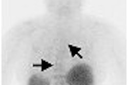
"Of the 206 patients reviewed, only three, or 1%, revealed metastases in the initially uninvolved extremities and/or brain," she said. "However, all three patients had widely metastatic disease with extensive visceral involvement."
Choi also noted that six studies showed new hypermetabolic foci in the same extremities involved by the primary melanoma. In addition, five patients had false-negative PET findings (one extremity and four brain lesions), and 11 false-positive PET extremity findings occurred in the cohort.
On the basis of the data reviewed by her research team, Choi suggested that melanoma patients should undergo FDG-PET/CT scans in the same manner as other oncology patients.
"The routine inclusion of the brain and the initially uninvolved extremities is of no significant benefit when compared with the information already obtained by the whole-body FDG study from the base of the skull to midthigh," Choi said. "Elimination of the extra scanning time may help to increase patient throughput and decrease radiation exposure for the patients."
By Jonathan S. Batchelor
AuntMinnie.com staff writer
March 30, 2006
Related Reading
PET/CT shows slight edge over MRI in melanoma staging, March 8, 2006
PET/CT shows potential for detecting unknown primary cancers, December 8, 2005
PET/CT imaging useful in detecting and staging choroidal melanomas, November 8, 2005
High-dose strontium-90 brachytherapy effective for posterior uveal melanoma, October 31, 2005
Oncologic PET/CT: A primer for radiologists, October 14, 2005
Copyright © 2006 AuntMinnie.com