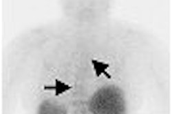
VIENNA - A recent prospective study comparing whole-body MRI with whole-body PET/CT in staging newly diagnosed malignant melanoma demonstrated that PET/CT has a slight edge in accuracy, but that both modalities missed local and distant metastases.
"A significant number of patients initially staged as N-negative and M-negative with whole-body MRI and PET/CT proved to have undetected regional or distant micrometastases, thus leading to low sensitivities for both modalities" said Dr. Florian Vogt from University Hospital Essen in Essen, Germany.
Vogt presented the results of research conducted by him and colleagues from the departments of diagnostic and interventional radiology, nuclear medicine, and dermatology at the European Congress of Radiology (ECR) this week.
"Malignant melanoma is the most aggressive skin tumor, and it frequently spreads to regional lymph nodes followed by organ metastases," he said.
Therapeutic options and the patient's prognosis strongly depend on the tumor stage, so accurate staging of the disease mandates a whole-body examination, according to Vogt. He and his fellow clinicians sought to compare the value of whole-body MR and PET/CT imaging in staging newly diagnosed malignant melanoma by correctly defining lymph node metastases (N) and detecting distant metastases (M).
Their cohort consisted of 31 patients with a mean age of 45 years who underwent a whole-body MRI and PET/CT after resection of the primary tumor. Because the tumor had been resected, its histology and Clark level were available to the researchers prior to the imaging exams.
The MRI exams were conducted on a 1.5-tesla Magnetom Sonata (Siemens Medical Solutions, Erlangen, Germany) equipped with an integrated phased-array surface-coil rolling table (BodySURF, MR-Innovation, Essen, Germany) capable of pulling the patient through the isocenter of the magnet.
The team conducted head-to-thigh scans with unenhanced T1 and T2 half-Fourier acquisition single-shot turbo spin-echo (HASTE) sequences of the liver and thorax. This was followed by seven overlapping contrast-enhanced axial 3D VIBE scans with a slice thickness of 3 mm, a matrix of 120 x 256, and an acquisition time of 22 seconds.
The PET/CT exams were performed on a Biograph (Siemens) system, with the patients imaged head to thigh. The studies were performed one hour after injection of 350 MBq of F-18 FDG, and CT was used for PET-based attenuation correction. A CT dataset was also acquired after the introduction of a negative oral contrast agent and the injection of 140 mL of iodine-based contrast, Vogt reported.
The MR images were read by two radiologists in consensus, while the PET/CT images were evaluated by a radiologist and nuclear medicine physician in consensus. M and N staging was conducted according to the 2003 American Joint Committee on Cancer (AJCC) classification.
"Histology and a clinical follow-up, with a mean of 295 days, served as standards of reference," he said.
The rates for sensitivity, specificity, positive predictive value (PPV), and negative predictive value (NPV) for N and M staging for both modalities are listed below:
|
||||||||||||||||||||||||||||||
Overall, the NM stage was correctly identified with whole-body MRI in 21 of the 31 patients (68%) and in 23 of 31 patients (74%) with PET/CT, Vogt reported.
"On the basis of these results for initial tumor staging, close follow-up examinations either by whole-body MRI or PET/CT seem to be mandatory to screen for recurrent disease," he said.
By Jonathan S. Batchelor
AuntMinnie.com staff writer
March 8, 2006
Related Reading
MRI, PET/CT show different strengths in tumor staging, January 20, 2006
PET/CT shows potential for detecting unknown primary cancers, December 8, 2005
PET/CT demonstrates high sensitivity in pulmonary nodule staging, December 2, 2005
PET/CT imaging useful in detecting and staging choroidal melanomas, November 8, 2005
Oncologic PET/CT: A primer for radiologists, October 14, 2005
Copyright © 2006 AuntMinnie.com