Pity the poor boy bander. JC Chasez from 'N Sync (no, not the one involved in the Super Bowl wardrobe malfunction) titled his 2004 solo effort "Schizophrenic" as a metaphor for the record's multiple musical stylings, i.e., personalities.
Chasez subsequently incurred the displeasure of the National Alliance for the Mentally Ill (NAMI), which called his use of the term schizophrenic "irresponsible" and "perpetuating misinformation" (the photo of Chasez in a straightjacket on the CD cover didn't help matters; the singer ultimately apologized).
But Chasez wasn't completely off the mark. The myth of multiple personalities aside, schizophrenia is a complex disorder. Specific diagnoses can range from paranoid schizophrenia (delusions about others) to catatonic (withdrawn from others) to undifferentiated (a little of both but not one more than the other). Then there are the other diseases -- bipolar disorder, Asperger's syndrome -- that are frequently misdiagnosed as schizophrenia.
Several recent studies have applied MRI to clarify schizophrenia. Three looked at whether imaging could offer details on the early stages of the disease; the fourth suggested that MR results can be the deciding factor between schizophrenia and bipolar disorder.
Corpus collosum
Abnormalities in the corpus collosum (CC) may be related to some of the symptoms and cognitive abnormalities that are seen in schizophrenia. But are these abnormalities already present in the early stages of the disease? U.K. researchers set out to answer this question with diffusion-tensor MRI.
"Diffusion-tensor imaging (DTI) ... appears to be sensitive to the presence of subtle white matter abnormalities in the absence of volumetric changes detectable by conventional MRI," wrote the group from the Institute of Neurology, University College London (Journal of Neurology, Neurosurgery, and Psychiatry, April 2005, Vol. 76:4, pp. 585-587).
In a previous DTI study, the group had successfully illustrated differences in the splenium of the CC of schizophrenics when compared with healthy subjects, suggesting focal disruption of commissural connectivity (JNNP, February 2000, Vol. 68:2, pp. 242-244).
Building on that research, the current study involved 20 patients with a DSM-IV diagnosis of schizophrenia (16 of whom were on antipsychotic medication) and 29 healthy volunteers. DTI was done on a 1.5-tesla scanner (Signa, GE Healthcare, Chalfont St. Giles, U.K.). The protocol included diffusion-weighted echo-planar imaging in the axial plane with the whole CC contained within 21 contiguous slices.
The results showed that there was no significant difference in mean diffusivity and fractional anisotropy between the two groups. The latter is a measure of white matter tract organization, while increased diffusivity may be linked to demyelination or axon loss.
However, "the present study failed to demonstrate abnormalities in the CC of patients with first-episode schizophrenia," the authors wrote. Reasons for this may include normal interhemispheric connectivity or pathological changes that are too subtle to detect in early stage illness. Instead, changes in the CC may be a result of chronic illness, they stated.
Frontostriatal function
"Understanding the pathophysiology of the emerging and progressing clinical manifestations is critical to understanding the pathogenesis of schizophrenia, and is likely to inform the development of preventive therapeutic strategies," wrote Aysenil Belger, Ph.D., in her group's study published in the Archives of General Psychiatry (March 2005, Vol. 62:3, pp. 254-262).
The researchers used functional MRI (fMRI) to identify potential frontostriatal dysfunction markers that may be linked to the pathophysiologic mechanisms of schizophrenia.
Belger is from the University of North Carolina, Chapel Hill; her co-authors are from Duke University in Durham, NC, and the University of Massachusetts Medical School in Waltham.
For this study 52 subjects were assigned to four groups: ultra-high-risk schizophrenia, early schizophrenia, chronic schizophrenia, and a control. The participants were presented with visual stimuli in a semirandomized sequential fashion.
During each imaging session, the subjects performed seven runs of a visual task during which they responded to various stimuli. A 1.5-tesla scanner (Signa, GE Healthcare) was used to acquire T1-weighted sagittal scans for coplanar anatomic images. Coplanar functional images were captured with a gradient-echo-planar sequence.
The areas of interest were the anterior cingulate gyrus (ACG), middle frontal gyrus (MFG), inferior frontal gyrus (IFG), the basal ganglia (BG), and the thalamus (TH).
"We hypothesized that, behaviorally, patients would demonstrate poor task-relevant target discrimination abilities relative to controls ... that prefrontal (MFG, IFG), medial frontal (ACG), and striatal (BG, TH) function ... would be diminished in all clinical groups," the authors stated.
The results proved their hypotheses true: the control subjects showed greater activation in prefrontal and medial frontal (ACG, IFG, MFG) areas than the clinical groups. Specifically, ACG activation was greater than MFG and IFG. They also found a decline trend of frontal and striatal activation across all four groups, indicating that prefrontal function decline begins before the onset of defined illness.
Ultimately, patients with schizophrenia demonstrated an inability to process relevant sensory information while simultaneously filtering out irrelevant input, the authors explained. These results dovetail with the dopamine hypothesis -- that schizophrenics suffer from dysregulated dopamine transmission, which leads to a stimulus-independent release of the neurochemical, creating inappropriate responses.
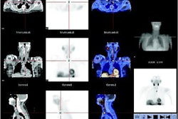
Using PET, another group from the National Institute of Mental Health (NIMH) also found a link between schizophrenia and dopamine. Dr. Andreas Meyer-Lindenberg, Ph.D., and colleagues took a closer look at the dorsolateral prefrontal cortex (DLPFC) and the hippocampal formation (HF). The group is from NIMH's Unit on Integrative Neuroimaging, as well as the Genes, Cognition, and Psychosis Program, both in Bethesda, MD.
The researchers measured cerebral blood flow in 22 patients with schizophrenia and 22 healthy controls. Imaging was performed, during a working memory challenge, on an Advance 3D scanner (GE Healthcare) after an injection of H215O per scan. They theorized that the most prominent disturbance of HF connectivity in schizophrenics would involve the link to the DLPFC.
According to their results, the patients showed reduced activation of the right DLPFC and left cerebellum during the working memory task. Additionally, inverse correlations between the HF and the contralateral DLPFC and the inferior parietal lobule were observed. Again, during the working memory task, the patients showed abnormal HF-DLPFC functional connectivity that persisted on through the control task. In comparison, this connection was diminished in healthy subjects.
"Our results are consistent with the possibility that the observed failure to modulate HF-DLPFC linkage and the lack of HF-DLPFC compartmentalization might to some degree contribute to the DLPFC dysfunction that is signature of (schizophrenia)," the authors explained (Archives of General Psychiatry, April 2005, Vol. 62:4, pp. 379-386).
This deficit in DLPFC circuitry, and subsequent dysfunction, is associated with dopaminergic disinhibition, they stated.
Hippocampal and ventricular volumes
In the final study, a multi-institutional team looked for significant neurological differences between schizophrenia patients and those with psychotic bipolar disorder (PBP) and nonpsychotic bipolar disorder (NPBP). "We hypothesized that the PBP and schizophrenia (patients) would resemble each other by having smaller hippocampal volumes and larger lateral + third ventricles than NPBP and (healthy controls)," wrote Dr. Godfrey Pearlson and colleagues (Biological Psychiatry, March 15, 2005, Vol. 57:6, pp. 633-639).
Pearlson and several of his co-authors are from the Yale University School of Medicine in New Haven, CT. Other contributors are from the Maryland Psychiatric Research Center in Catonsville, MD; Johns Hopkins University School of Medicine in Baltimore; Brigham Young University in Provo, UT; and LDS Hospital in Salt Lake City.
The patient population consisted of 38 BP subjects, 23 of whom had psychosis, defined as the occurrence of hallucinations and/or delusions during at least one affective episode. There were also 33 patients with schizophrenia and 44 healthy subjects.
All MR scans were obtained on a 1.5-tesla scanner (Signa, GE Healthcare). Contiguous coronal slices with spoiled gradient-echo and steady-state sequences were acquired. Volumetric measurements were generated with MEASURE, a custom graphics software program developed by co-author Patrick Barta, Ph.D.
According to the results, no significant group difference was seen for left hippocampal volume, although it tended to be larger for the NPBP group than for the control. The authors found this surprising because previous research had implicated the hippocampus in BP.
However, PBP and schizophrenia patients had significantly enlarged lateral and third ventricles when compared with the control and NPBP, suggesting that PBP and schizophrenia can overlap, while NPBP is a different type of disorder.
"This study … is important because it is the first clear demonstration of anatomic differences in PBP but not in NPBP that resemble those seen in schizophrenia," the authors concluded. Making these distinctions is vital for determining treatment protocols and their effect on brain structure, they added.
By Shalmali PalAuntMinnie.com staff writer
April 27, 2005
Related Reading
AuntMinnieTV: DTI offers insight into ADHD, January 4, 2005
Olanzapine has various effects on striatal volumes in schizophrenia, October 26, 2004
Susceptibility-weighted MRI ups contrast, offers minute detail, September 15, 2004
Copyright © 2005 AuntMinnie.com