"If one meditates on the Medicine Buddha, one will eventually attain enlightenment, but in the meantime one will experience an increase in healing powers both for oneself and others, and a decrease in physical and mental illness and suffering."
Mindful meditation is often described as the ethereal state between wakefulness and unconsciousness. Those who meditate regularly -- with Buddhist Rainbow Light Meditation, Kundalini yoga, or Christian centering prayer -- talk of physical calm, mental peace, and a pervasive sense of well-being.
Now imagine trying to achieve that kind of bliss as an MR scanner drones around your head, or a radioactive agent is pumped into your veins through an IV -- all while a bunch of scientists keep an eagle eye on your brain's every move.
Fortunately, there are hard-core meditation buffs who can manage mind over matter, making them ideal for neuroimaging studies. But monks, yogis, and gurus aren’t the only ones benefiting from meditation. As modern life becomes increasingly stressful, regular people are grasping for ways to cope. Ten million adults in the U.S. now practice some form of meditation, whether sponsored by an employer, sanctioned by a school during recess, or done on the fly in an airport lounge (Time, August 4, 2003).
In turn, the mental health community is taking a closer look at the effects of meditation on the brain and its function. Many of these scientists will gather this weekend at the Massachusetts Institute of Technology in Cambridge for "Mind & Life XI: Investigating the Mind: Exchanges between Buddhism and the Biobehavioral Sciences on How the Mind Works."
The Dalai Lama, considered the religious and political leader of Tibet, is scheduled to participate in the two-day seminar. Other participants include Nancy Kanwisher, Ph.D., from the McGovern Institute for Brain Research at MIT, whose research has used fMRI and magnetoencephalography (MEG) to investigate visual object recognition, and Stephen Kosslyn, Ph.D., who has worked with PET and functional MRI (fMRI) to study the nature of visual mental imagery. Kosslyn is from Harvard University in Boston.
Kosslyn will take part in a "Mind & Life" session on mental imagery, delving into "the relationship between the conscious experience and the neural mechanisms of mental imagery," he wrote in an email to AuntMinnie.com. Kosslyn said his talk would focus on the mechanics of introspection and inner perception.
 |
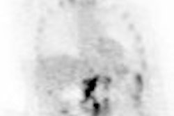
Above and below, images from a 2.5-second movie shot during a session of practice of
transcendental meditation, using a 148-channel machine. This head montage shows a topographic display of MEG of the raw data upper right (graph of 148 superimposed images). This research was done collaboratively by the Brain Research Institute of Fairfield, IA and the Neuromagnetic Laboratory, Henry Ford Hospital, Detroit. Images courtesy of Alarik Arenander, Ph.D., Brain Research Institute.
  |
Another speaker will be Dr. Richard Davidson, Ph.D., from the University of Wisconsin in Madison. Davidson is considered a pundit of meditation imaging, having convinced Tibetan monks from Dharamsala, India to undergo both MR and PET brain scans. So far, the gist of Davidson’s ongoing research is that, thanks to routine meditation, these monks maintain warm, fuzzy feelings continuously (i.e. their prefrontal lobes, the brain’s area associated with positive emotions, light up like crazy, even when they aren’t meditating).

"So an interesting question for neuroscientists is how do the brains of Buddhist practitioners -- or indeed any other wise, happy and virtuous people -- light up? How are the qualities of happiness, serenity and loving kindness that arise from the Buddhist practice of mindful meditation reflected in the brain? How does that subjective experience manifest itself?" asked philosopher Owen Flanagan, Ph.D., in an essay called "The colour of happiness" (New Scientist, May 24, 2003, Vol. 178:2396, pp. 44).
Relying on a variety of imaging techniques, investigators are seeking to enlighten all of us about the objective virtues of a contemplative life.
SPECTacular concentration
As strenuous as it may be to maintain serenity during an imaging exam, taking a closer look at meditation practices is just as challenging for clinicians, pointed out Dr. Andrew Newberg from the division of nuclear medicine at the University of Pennsylvania Medical Center in Philadelphia.
"The study of meditation requires us to push the imaging technologies to their limits in many ways," Newberg said. "It is difficult to assess the subjective nature of the meditative experiences, and how that correlates with cerebral function. It challenges design paradigms, and it informs us about the network of cerebral interactions that occur during complex tasks."
Newberg would know, having led several studies that used SPECT imaging on meditators.
In one such study, his group analyzed 99mTC-HMPAO SPECT data from eight veteran practitioners (15 years or more) of Tibetan Buddhism. The SPECT scans measured regional cerebral blood flow (rCBF), which correlates closely with cerebral activity, Newberg and co-authors wrote (Psychiatry Research: Neuroimaging, April 10, 2001, Vol. 106:2, pp. 113-122).
In order to compare meditative changes to baseline, the subjects underwent imaging twice on a triple-head gamma camera using high-resolution fan-beam collimators (Prism, Picker International). They were injected with 7 mCi of 99mTC-HMPAO for the first 45-minute scan, and with 25 mCi of 99mTC-HMPAO for the second 30-minute scan.
Before the second scan, "the subject meditated for approximately one hour at which time the subject provided a ‘signal,’ observable to the investigators, that was incorporated as part of the meditation practice," they explained.
The rCBF was measured in selected regions of interest (ROI) including the inferior frontal, superior frontal, dorsolateral pre-frontal, cerebellum, and cingulate gyrus.
 |
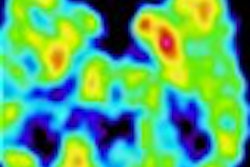
| The images show the results from a baseline SPECT scan on the left (i.e. at rest) and during a "peak" of meditation shown on the right. Explained Dr. Andrew Newberg: "Two sets of images were taken, showing slightly different parts of the brain. The first image shows decreased activity in the parietal lobe (lower right shows up as yellow rather than the red on the left image) during meditation. This area of the brain is responsible for giving us a sense of our orientation in space and time. We hypothesized that blocking all sensory and cognitive input into this area during meditation is associated with the sense of no space and no time, which is so often described in meditation." |
According to the results, relative increases in rCBF were observed in several areas, such as the inferior and orbital frontal cortices, the sensorimotor cortices, the midbrain, and the thalami. The change in activity in the left dorsolateral pre-frontal cortex (DLPFC) correlated positively with the change in activity in the left thalamus, as well as positively with a change in activity in the right thalamus, and negatively with the change in activity in the left superior parietal lobe.
 |
| This image shows that the front part of the brain, which is usually involved in focusing attention and concentration, is more active during meditation (increased red activity). This makes sense as meditation requires a high degree of concentration. Newberg wrote: "We also found that the more activity increased in the frontal lobe, the more activity decreased in the parietal lobe. A more complex version of the model from which the hypothesis is based can be found in the book Why God Won’t Go Away: Brain Science and the Biology of Belief by Andrew Newberg, Eugene d’Aquili, and Vince Rause, Ballatine Books, New York, 2001. |
Some of the disorders related to the DLPFC are depression, lack of motivation, and memory problems. Elaborating on the clinical significance of these findings, Newberg wrote in an e-mail to AuntMinnie.com that this type of information could provide a theoretical framework for future studies.

"This includes studies attempting to explore the relationship between neurotransmitters and meditation, since the effects on serotonin or dopamine may very well have implications for the use of meditation in disorders such as depression, attention deficit disorder, and/or Parkinson’s disease," he wrote.
Results from Newberg’s latest study are set for a forthcoming issue of Perceptual and Motor Skills. The researchers will look at cerebral blood flow during meditative prayers in the same group of Tibetan Buddhists, as well as in centering prayer, performed by Franciscan nuns. One of the main purposes of the study is to show that neuroimaging techniques can be applied to different types of meditation.
Never mind the magnet
One reason Newberg said he chose SPECT over MRI was his concern that noise from the magnet would be too distracting. But other researchers have successfully relied on fMRI. One such study, spearheaded by the Mind-Body Medical Institute in Boston, used MR for functional brain mapping of meditation and the relaxation response.
Lead by Sara Lazar, Ph.D., from Massachusetts General Hospital in Boston, the investigators chose fMRI because of its ability "to identify foci of activity that are modulated by a very simple form of meditation" (NeuroReport, May 15, 2000, Vol. 11:7, pp. 1581-1585).
The group recruited five subjects who had practiced Kundalini meditation daily for at least four years. This version of Kundalini required the participants to passively observe their breath and silently repeat Sanskrit phrases during inhalations and exhalations. Prior to the study, the participants were given an audiotape of the sound of the beeping scanner, and asked to practice meditating with the tape running.

The subjects underwent imaging on a 3-tesla scanner (GE Medical Systems, Waukesha, WI) at MGH’s NMR Center. Sixteen 7-mm gradient-echo functional slices were obtained using a quadrature head coil. Activity during the last six minutes of each meditation period was compared to the data from six-minute control periods. Late versus early meditation were also compared, according to the authors.
The results showed significant increases in fMRI signal during meditation in the following brain regions: putamen, midbrain, pregenual anterior cingulate cortex, and hippocampal/parahippocampal formation. This increased pattern of activity was maintained throughout two scanning sessions, with greater activation in the foci and a larger percentage signal change during the second scan.
"These fMRI signal increases were robust...these findings suggest that neural activity during meditation is dynamic, slowly evolving during practice," they concluded. Ultimately, studies such as this should make the practice of meditation more palatable as a form of complimentary medicine.
Meanwhile, Davidson’s continuing work at the Keck Laboratory for Functional Brain Imaging and Behavior at Wisconsin has revealed that the persistent activity in the left prefrontal lobes (associated with mood, emotion, and temperament) may override activity in the right prefrontal lobe that contributes to that familiar lousy feeling. In addition, meditation also may be able to help control the response of the amygdala, leading to less fear, anger, and anxiety.
 |
| The main temple at Dharamsala in India, the home of many Tibetan monks and nuns in exile. |
A study from the University of Pennsylvania in Philadelphia attests to the importance of keeping the amygdala in check. Using event-related fMRI, the researchers tested their hypothesis that voluntary modulation of negative emotion is associated with changes in neural activity. The subjects were shown negative (a pointed gun) and neutral (a rolling pin) images. They were then asked to either maintain their initial emotional response to the image, or passively view the photo without any self-modulation. The researchers found a prolonged fMRI increase in signal change in the amygdala when the subjects were asked to maintain the negative emotional response.
"These results suggest that consciously evoked cognitive mechanisms that alter the emotional response...(alter) the degree of neural activity in the amygdala ... increased amygdalar activity is associated with symptom provocation in anxiety disorders (and) may be the cause of unregulated negative effect observed in (depression)," the authors explained (Journal of Cognitive Neuroscience, August 15, 2002, Vol. 14:6, pp. 913-921).
Others are experimenting with different modalities. A group from the University of Graz in Austria imaged two QiGong experts with transcranial Doppler sonography (TCD), and near-infrared spectroscopy, which measures oxygenation levels in cerebral tissue.
The goal of the study was to "clarify some of the old mysticisms of this Chinese meditation exercise...with new brain function monitoring," explained lead author Dr. Gerhard Litscher (Neurological Research, July 2001, Vol. 23:5, pp. 501-505).
The group found that during meditation, the mean blood-flow velocity on TCD increased in the right posterior cerebral artery and decreased in the left middle cerebral artery. The spectroscopy scan showed a simultaneous increase in oxyhemoglobin and total hemoglobin. Finally, "during QiGong EEG alpha activity occurred predominantly in the anterior half, and occurred silently in the posterior half of the brain." This cerebral ying and yang shows that "QiGong is...a special state of excitation, not a state of consciousness between wakefulness and sleep," thereby putting a different twist on the meditative state.
Peace of mind for the masses
Regardless of the imaging modality used, one element that these studies have in common is that experienced practitioners of meditation were involved. Would the results be different for any old Joe/Jane Office Worker who meditated daily at his or her desk for a few minutes?
A paper in Psychosomatic Medicine by Davidson’s group gathered EEG data on 48 employees of a Madison-based biotechnology company who had gone through an eight-week training program in mindful meditation, as well as an influenza vaccination. Specifically, they were looking for alterations in the brain and immune system post-meditation.
They came up with "significant increases in the left-sided anterior activation, a pattern previously associated with positive effect, in the meditators compared with the nonmeditators. We also found significant increases in antibody titers to influenza vaccine among subjects in the meditation (group)" (Psychosomatic Medicine, July-August 2003, Vol. 65:4, pp. 564-570).
Studies on meditation newbies are particularly important to answer a classic chicken-or-egg quandary: Does lifelong meditation bring about a change in brain function, or are some people simply neurologically wired for penitence?
"Larger trials that include imaging and people of various levels of practice along the way would need to be studied. It is certainly an expensive and lengthy study to perform, but I think that the results could be very important," Newberg commented.
 |
| In May 2001, Tenzin Gyatso, the 14th Dalai Lama of Tibet, toured the Keck Laboratory for Functional Brain Imaging and Behavior at the University of Wisconsin. Here, he takes a gander at the facility’s PET scanner. Photo by Jeff Miller. Image courtesy of the University of Wisconsin. |
In the meantime, there’s still plenty of information to be gleaned from the great meditators of our time. So if Newberg could recruit anyone he wanted for a meditation study, whom would he choose? The man revered by his followers as the 14th reincarnation of the Buddha: the Dalai Lama.
"Theoretically speaking, individuals such as the Dalai Lama are perpetually in an enlightened state and therefore may not show as much change during meditation as a more novice meditator," Newberg explained. "On the other hand, if one wanted to capture the most profound states achievable, then the Dalai Lama might be the best person. As far as the imaging modality to use, I think the best type of study would cross compare several modalities at the same time such as EEG, fMRI, and perhaps a neurotransmitter ligand for PET. This would allow us to capture many different cerebral processes simultaneously."
By Shalmali PalAuntMinnie.com staff writer
September 11, 2003
Related Reading
Meditation shown to light up brains of Buddhists, May 23, 2003
Strike a pose: PET sheds light on benefits of yoga, December 31, 2002
Copyright © 2003 AuntMinnie.com