SAN DIEGO - Mammography and breast ultrasound are the most commonly used modalities for breast cancer screening. However, a positive mammographic finding does not always correspond to a malignancy. FDG-PET has shown great utility in differentiating benign from malignant lesions in invasive ductal carcinoma, and can also be used in patients with inconclusive findings from conventional imaging modalities.
A group of researchers from the departments of surgery, radiology, and pathology at Wake Forest University School of Medicine in Winston-Salem, NC, have conducted a preliminary study of functional breast imaging of positron emitters coupled with mammography. The co-registration technology, dubbed positron emission mammography (PEM) and developed by PEM Technologies of Bethesda, MD, shows promise for enhanced cancer detection, biopsy guidance, and the reduction of unnecessary biopsies of mammographically suspicious benign lesions.
In a presentation at the 2002 Academy of Molecular Imaging conference last week, Dr. Irving Weinberg, president of PEM Technologies, outlined the results of the researcher’s investigations with the new technology.
The study was performed using PEM in 16 women scheduled for stereotactic breast biopsy for additional evaluation of abnormal mammograms. The 16 patients, all over 18 years of age, were recruited from the university’s surgical clinic, and their consent was obtained for the investigational procedure.
The patients fasted for four hours, and were then injected with 10 mCi of FDG. Imaging began one hour later. The scans were performed over 4 minutes without releasing compression by a dual-head bismuth germanate oxide (BGO) PET scanner, and a limited-angle iterative reconstruction of the images was conducted. With the breast in the same position, the stereotactic imaging and biopsy were performed.
"The core biopsy was done solely under stereotactic x-ray, as this could not be done using the PEM system due to its status as an investigational device," Weinberg said.
The images were then reviewed and compared to pathologic results. The team co-registered the x-ray and the PEM images and obtained the 3-D location of the biopsy site on the PEM images. A total of 18 lesions in 17 breasts were evaluated, and 7 cancers were found.
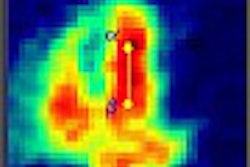
The PEM images were analyzed by drawing a region of interest (ROI) at the biopsy site and comparing the mean count density in the biopsy ROI with the mean count density of a background ROI in the same z-plane as the biopsy, Weinberg said. The group determined that a lesion-to-background ratio of 2.5 would be a robust indicator of malignancy for this initial study.
The threshold resulted in a sensitivity of 86%, a specificity of 91%, and an overall diagnostic accuracy of 89% with the use of PEM. The accuracy with the use of only x-ray resulted in a diagnostic accuracy of 39%. In regards to tumor size, the results of the PEM study corresponding to pathology were 6 mm with PEM and 7 mm with pathology, 7 mm and 6.5 mm, 8 mm and 10 mm, and 30 mm and 30 mm, respectively. This resulted in a 0.97 correlation with PEM to pathology.
The technology is seen as aiding presurgical planning and guiding biopsies, as well as reducing unnecessary biopsies. Its technical goals are to determine cancer size and the presence of additional foci, with the foci targeted for biopsy with x-ray correlation. The technique can also be used to characterize tumors as benign or malignant, Weinberg said.
"This initial clinical trial with PEM and x-ray shows great promise for its capabilities to assist radiologists and surgeons with staging and treatment for invasive ductal carcinoma," he said.
The company plans on developing the next generation of its PEM technology (PEM 2400) with lutetium oxyorthosilicate (LSO) crystals in order to achieve a higher effective number of protons per atom and density compared to BGO, resulting in a higher detection efficiency. In addition, the use of LSO crystals will provide a short decay constant for good coincidence timing and a higher light output, Weinberg said.
By Jonathan S. BatchelorAuntMinnie.com staff writer
October 28, 2002
Related Reading
Scintimammography catches chemoresistance in breast cancer patients, June 13, 2002
FDG-PET predicts outcome in post-therapy breast cancer patients, March 8, 2002
PET offers important benefits in breast cancer imaging, October 29, 2001
Copyright © 2002 AuntMinnie.com