Tuberculosis (TB) is the single largest cause for pericarditis in India. Nearly 50% of the pericarditis cases develop pericardial constriction. This in turn can lead to abscess formation requiring speedy diagnosis and intervention. However, not many studies are available on the subject despite its prevalence in India and other developing countries.
Researchers from the premier medical institution in India, the All India Institute of Medical Sciences, attempted to close this gap with a study of 13 patients who developed pericardial abscess from a group of 120 with pericardial constriction. The study used both CT and MRI to determine the location and the extent of the abscesses with respect to the surrounding cardiovascular structures.
CT was used in 11 cases and MRI in seven cases. Five of the 13 patients underwent both CT and MRI. And at the end of the study, published in Clinical Radiology, the authors, Dr Gulati G.S and Dr Sharma S, found 15 abscesses and concluded, "The ability of CT and MRI to demonstrate the anatomic details of the pericardium consistently and accurately makes them invaluable for the assessment and management of patients with focal or generalised pericardial lesions" (Clinical Radiology, June 2004, Vol. 59:6, pp 514-519).
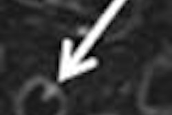
CT was performed on a Picker CT scanner (now Philips Medical Systems, Andover, MA, U.S.). Non-contrast scanning was done with 8-10-mm-thick axial sections from the lung apices to the domes of the diaphragm, followed by contrast-enhanced scans using nonionic contrast (iohexol 360 mg/ml; 2 ml/KBW) with a delay of 8-12 sec. Thinner 4-mm sections were taken where lesions were present along the right and left margins of the heart.
The authors observed that the lesions had a smooth and thin enhancing rim in 91% and 54% of cases, respectively. They noted calcification in 27% of cases. All of the lesions had a hypodense core.
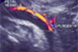
MRI was done using a Magnetom 1.5-tesla unit (Siemens Medical Systems, Erlangen, Germany). Acquisitions were ECG-gated and performed in axial, sagittal, and coronal planes. The slice thickness was 5 mm, and the interslice interval was 1-2 mm.
"Spin-echo (SE) T1-weighted images were acquired with a TR at 80% of the R-R interval, minimum TE, minimum field of view without incurring wraparound artifacts. SE T2-weighted images were acquired with a TR of about 3-4 times the R-R interval, TE of approximately 80 ms, 256 x 128 matrix, and two signal averages," the authors wrote.
The MRI showed four patients, or 57% of the patient group, had hypointense core, and three patients had an isointense core on SE T1-weighted images. On the T2-weighted and gradient-echo (GRE) images, five patients, or 71% of the patient group, had a hyperintense core.
Interestingly, one patient had a hypointense core on the T2-weighted and GRE image. "This unusual finding for tuberculous involvement of the pericardium has not been previously reported. Presence of hypointensity on T2-weighted images may suggest tuberculosis," the authors noted.
However, the authors summarised that MRI was useful in evaluating the location and extent of the disease, but lacked disease-specific image morphology. And that both CT and MRI were superior in the evaluation, localization, and extent of lesions not in contact with the chest wall, compared to echocardiography, which is limited by poor acoustic window.
The imaging findings from the study show that predominant involvement of the right atrioventricular groove with features suggesting localised pericardial tamponade should warn clinicians and radiologists to the likelihood of pericardial abscess.
The results represent a valuable input to the diagnosis and treatment of TB-related complications, especially in countries where the disease continues to be a major threat.
By N. ShivapriyaAuntMinnieIndia.com staff writer
July 21, 2004
Copyright © 2004 AuntMinnie.com