Older patients need less contrast than younger adults for pancreatobiliary imaging, researchers from Japan report. The results of their study, published in the August American Journal of Roentgenology, showed that a 12% contrast dose reduction by body weight produced the optimal contrast profile in multidetector-row CT's triphasic imaging protocols.
The controllable factors in contrast-enhanced CT imaging of the abdomen have a substantial effect on its diagnostic capabilities, wrote the researchers from the Nagoya University School of Health Sciences and Graduate School of Medicine in Nagoya, Japan. But what has been considered optimal enhancement was too intense in the pancreas.
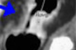
"Previous studies have reported that to evaluate anatomic structures and various pathologic conditions of the pancreatobiliary region on contrast-enhanced CT, it is essential to acquire pancreatic-phase images showing intense enhancement of the pancreatic parenchyma," wrote Drs. Shigeki Itoh, Mitsuru Ikeda, Hiroko Satake, and colleagues. "However, when interpreting CT images acquired using that protocol in our clinical practice, we noted that in some examinations of elderly patients, because contrast enhancement of the pancreatic parenchyma in pancreatic-phase images was too intense, the standard settings for window width (345 HU) and level (45 HU) used at our institution had to be adjusted to evaluate the pancreatic parenchyma in detail" (AJR, August 2006, Vol. 187:2, pp. 505-510).
The study sought to determine whether it was possible to reduce the contrast dose and injection rate in elderly patients undergoing arterial, pancreatic, and portal venous phase CT of the pancreatobiliary region with MDCT.
The researchers divided 112 patients (74 men, 38 women; ages 37-80; mean body weight 57 kg) into three groups:
- Group 1 - 49 patients 60 years or younger, 0.08 mL kg body weight (up to 5 mL/sec)
- Group 2 - 32 patients > 60 years of age, 0.08 mL kg body weight (up to 5 mL/sec)
- Group 3 - 31 patients > 60 years of age, 0.07 mL kg body weight (up to 4.5 mL/sec)
Patients were instructed to drink 300 mL of water five minutes before scanning to opacify the gastrointestinal tract. Nonionic contrast material (concentration 300 mgI/mL) was injected for 30 seconds, followed immediately by a 5% dextrose flush at 5 mL/s over six seconds. Images were acquired on a 16-detector scanner (Aquilion, Toshiba Medical Systems, Tokyo). Arterial and pancreatic-phase images were acquired at 16 x 0.5-mm collimation and 450 mAs. Portal images were acquired at 16 x 1-mm collimation and 400 mAs.
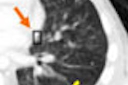
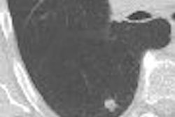
Two radiologists blinded to the patients' clinical information and contrast protocol graded the degree of contrast enhancement on a five-point scale, and also assessed attenuation quantitatively for enhanced and unenhanced scans on a workstation in circular regions of interest at the aorta (superior, middle inferior), portal vein, splenic vein, superior mesenteric vein, pancreas, and liver.
The results showed no significant differences between the groups in the mean start time of arterial-phase scanning. However, compared with groups 1 and 2, "group 3 achieved a 12.2% reduction in the volume and rate of contrast injection," the researchers wrote.
"Contrast enhancement in the main phases for all organs was significantly more intense in group 2 (full dose, age over 60) than in groups 1 and 3," they wrote. "Cases in which pancreatic enhancement in the pancreatic phase was graded as excessive were more frequently observed in group 2. No statistically significant differences were observed between groups 1 and 3 in either quantitative or visual assessment for enhancement of any organ in any phase."
"The results ... have shown that compared with patients 60 years old and younger, it is possible to achieve a 12.2% reduction in the volume and rate of contrast material injection in patients more than 60 years old, while maintaining equivalent contrast in terms of both quantitative and visual assessment on contrast-enhanced CT studies of the hepatobiliary region," Itoh and colleagues wrote.
The diminished need for contrast is due to several factors, including a reduction in cardiac output and blood volume, commonly seen in elderly patients, that aids abdominal contrast by delivering a better bolus.
"Previous studies have also shown that contrast enhancement during the early phase after the injection of contrast material is inversely related to cardiac output and blood volume," the group wrote. They added that contrast material tends to be more slowly excreted in patients with reduced cardiac output, which leads to better enhancement in the late phase.
Finally, they noted, the circulatory dynamics of the pancreas, "a well-perfused organ with an exclusively arterial blood supply from large arteries in the abdomen," can render enhancement that is too intense for standard window settings if the dose and rate of contrast enhancement are not reduced.
As for limitations, the upper limits on contrast suggest that further reductions in contrast enhancement are possible, especially in the oldest patients. Moreover, the cohort was small and more study is needed to assess contrast needs in specific pathologic conditions.
The dose and rate of contrast should be reduced by at least 10% in patients older than 60, the researchers concluded. "It should be noted that this helps to reduce both the cost of the examination and the risk of complications," they wrote
By Eric Barnes
AuntMinnie.com staff writer
August 2, 2006
Related Reading
N-acetylcysteine reduces incidence of contrast-induced nephropathy June 29, 2006
MDCT makes inroads in urology, biopsy guidance, and oncology imaging, June 19, 2006
Concentrated iodine permits lower volumes in cardiac CTA, May 22, 2006
The more CT detectors, the more MPRs help, March 22, 2006
MDCT yields reliable diagnosis of pancreas divisum, April 14, 2005
Copyright © 2006 AuntMinnie.com