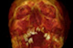
ORLANDO, FL - To the surprise of investigators, CT scans of mummies at the Egyptian Museum in Cairo show that these ancient Egyptians had signs of atherosclerosis in their hearts and arteries.
"We really didn't expect to find any atherosclerosis in these mummies," said Dr. Samuel Wann, chair of the department of cardiovascular medicine at the Wisconsin Heart Hospital in Wauwatosa.
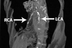
However, he and his fellow researchers from the U.S. and Egypt discovered calcification in nine of 16 mummies in which cardiac organs or vascular structures were identifiable.
 |
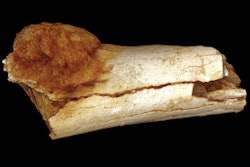
| All images courtesy of Dr. Michael Miyamoto of the University of California, San Diego. |
"The calcification we saw in the CT scans was exactly what we see in people today," said Dr. Randall Thompson, professor of medicine at the Mid America Heart Institute in Kansas City, MO, in a presentation at this week's American Heart Association (AHA) conference.
The researchers examined 33 mummies of people who died as long as 3,500 years ago. Thompson suggested that because mummies represent elite classes -- people who worked in the royal household, royalty themselves, and, in one case, a soldier -- they were upper-middle-class individuals, who apparently were susceptible to the same diseases as modern society.
 |
The state of decomposition of the mummies made it difficult to determine the exact cause of death, said Thompson. Seven or eight mummies, identified as individuals who were older than 45 years of age, showed signs of arteriosclerosis.
The researchers worked with curators and doctors at the Cairo museum. The mummies were taken from the museum to a 64-detector-row CT scanner (Somatom Emotion 6, Siemens Healthcare, Erlangen, Germany) on a trailer behind the facility, where they were scanned. They were not unwrapped, although some of the mummies had previously had their heads exposed.
The mummies dated from 1981 B.C. to 364 A.D. Social status could be determined for most of them -- all were of high status. The researchers found no difference in calcification between men and women, and the most ancient mummy with findings diagnostic of atherosclerosis died between 1530 and 1570 B.C.
By Ed Susman
AuntMinnie.com contributing writer
November 18, 2009
Related Reading
Austrian study uses MDCT to uncover tale of 12 mummies, October 5, 2009
CT reveals Nefertiti's secrets, April 1, 2009
Philips CT images Egyptian mummy, February 5, 2009
Scan artist: Radiologist uses CT to reveal mystery of antiquities, October 25, 2005
CT helps unwrap mummy mystery, March 29, 2005
Copyright © 2009 AuntMinnie.com