A compact but concentrated dose of high-osmolarity contrast agent produced a better MR contrast profile than gadolinium in patients with chronic thromboembolic pulmonary hypertension, a U.K. team has reported.
Chronic thromboembolic pulmonary hypertension (CTEPH) occurs in a small subset of patients following acute embolism, and results from incomplete clot lysis that subsequently organizes and adheres to the vessel wall.
Better visualization of vessels can only help matters in a condition like CTEPH, which often remains undiagnosed. Typical progressive dyspnea and exercise intolerance are often attributed to other conditions, though treatment is effective. Recanalization can at least partially restore blood flow through the obstructed vessel. Eventually, untreated CTEPH can lead to progressive pulmonary hypertension, right ventricular failure, and death.
"Previous studies have compared 0.5 M and 1.0 M contrast agents in the evaluation of pulmonary vasculature and found little or no benefit when SNR (signal-to-noise ratio) was compared between the two agents," wrote Drs. Neil Woodhouse, Jim Wild, Edwin van Beek, and colleagues from the University of Sheffield and Sheffield Teaching Hospitals NHS Trust in the U.K. and the University of Iowa in Iowa City. "However, when high-molarity contrast agents have been evaluated for MRA (MR angiography) of the pelvis, increased SNR and better small vessel opacification were demonstrated."
Their research, presented as a poster at this month's International Society for Magnetic Resonance in Medicine (ISMRM) conference in Seattle, compared gadolinium (Gd-DTPA, Magnevist, Schering, Berlin) to Schering's high-molarity Gd-BT-D30A (Gadovist) contrast agent.
The exams were performed on six patients twice in 48 hours (one with each contrast agent in random order) using a 1.5-tesla whole-body MR system (Eclipse, Philips Medical Systems, Andover, MA).
"The high-resolution scans were acquired during 20 seconds of arrested inspiration after a period of hyperventilation using a coronal 3D RF-spoiled gradient-echo sequence (TR/TE 5 msec/2 msec, matrix 256 x 440, FOV [field-of-view] 400 mm, slice thickness 2 mm, flip angle 50º 1 NEX)," the group explained.
A small test bolus was used to assess contrast transit time, followed by a saline flush, and injection delays were set to ensure that the center of the k space coincided with a bolus peak. The contrast and saline were administered via power injector (Spectris, Medrad, Indianola, PA) at 0.2 mmol/kg Gd-DTPA (or Gd-BT-DO3A) plus 20 mL saline at 5 mL/sec.
Time-resolved MRA was acquired during a 22-second breath-hold with coronal 3D RF-spoiled gradient-echo sequence at the rate of 1 frame/2.5 fps (TR/TE 3 msec/2 msec, matrix 64 x 256, FOV 450 mm, slice thickness 10 mm, flip angle 30°, 1 NEX). Half the contrast dose was administered (0.1 mmol/kg Gd-DTPA or Gd BT-DO3A) followed by 15 ml saline.
For image analysis, the group first chose regions of interest (ROI) in the coronary arteries up to the fifth order of branching, with ROIs comprising the entire vessel.
"Bolus profiles were derived from an ROI placed in the main pulmonary artery and another in the aorta on a single slice from the dynamic time-resolved series of images," the group wrote. Each slice was also examined parametrically with Matlab code, in addition to visual review by a radiologist blinded to the contrast type.
The results showed a far greater separation of pulmonary arterial and aortic peaks in the Gd-BT-DO3A examinations when compared to the Gd-DTPA, with a faster contrast washout. On the images, this produced less image contamination due to cardiac motion, cardiac and pulmonary venous and systemic vasculature.
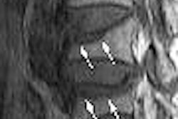
"The mean contrast-to-noise ratio in all orders of a vessel was higher for Gd-BT-DO3A," Woodhouse and colleagues wrote. "Filling defects and intraluminal webs were better visualized. In five out of six patients, a higher order of vasculature was resolved with Gd-BT-DO3A."
Finally, a comparison of pixel-to-pixel parametric maps between the two time-resolved exams showed better separation of pulmonary arterial and venous circulation with the new agent, they stated.
"Pulmonary angiography is ultimately dependent upon the cardiac output of the right side of the heart (especially in CTEPH), and a smaller volume results in less 'smearing' of the bolus geometry," the group wrote.
The study found that Gd-BT-DO3A's more compact bolus profile yielded less contamination of the arterial-phase image by pulmonary venous and systemic vasculature, and therefore less image degradation due to cardiac motion. High-osmolar contrast improves the visualization of smaller vessels, while facilitating the separation of arterial and venous circulation, the group concluded.
By Eric Barnes
AuntMinnie.com staff writer
May 30, 2006
Related Reading
Perfusion MRI: A multifaceted modality for myocardial ischemia, May 8, 2006
MR angiography offers greater detail in vascular imaging, March 10, 2006
High resolution, fast acquisition give 3D MRA edge in abdomen, January 20, 2006
ACC/AHA issue new treatment guidelines for peripheral artery disease, December 7, 2005
Copyright © 2006 AuntMinnie.com