ATLANTA - Stress perfusion defects found with PET/CT are good predictors of coronary artery disease, while only overall perfusion defect changes are good predictors of cardiovascular events with PET, according to two studies presented at the American College of Cardiology (ACC) meeting.
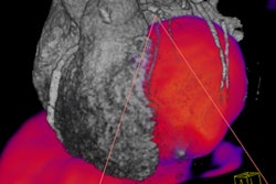
In the first study, 64 patients with suspected coronary artery disease (CAD) and 38 patients with low likelihood of CAD underwent rest-stress rubidium-82 cardiac PET/CT and coronary angiography. The tests were performed no more than six months apart.
Stress perfusion defects found with PET/CT detected 41 of the 44 cases of obstructive CAD found with angiography, a sensitivity of 93%. Compared to angiography, PET/CT had an overall diagnostic accuracy of 87% and a specificity of 83% for excluding obstructive CAD.
Sensitivity was somewhat higher for multivessel compared to single-vessel disease (95% versus 92%).
PET/CT's sensitivity for "detecting single-vessel disease and the extent of multivessel CAD is higher than that reported for any other technique," according to lead author Dr. Uchechukwu Sampson, a research fellow at Vanderbilt University in Nashville, TN.
All the patients with low likelihood of CAD had normal PET/CT scans. Overall sensitivity was similar between men and women, as well as between obese and nonobese patients.
However, Sampson cautioned that cardiac PET/CT may underestimate the anatomic extent of CAD in some patients.
In the second study, researchers found that, when using PET to predict future coronary events in patients under therapeutic management, it may be more effective to track overall myocardial perfusion changes in the entire coronary vascular tree rather than regional changes, such as at the site of the most severe coronary lesion.
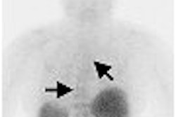
A team of researchers led by Dr. Stefano Sdringola, a cardiologist at the University of Texas at Houston, examined 409 patients with CAD using N-13 ammonia PET with dipyridamole pharmacologic stress initially and after 1.4 to 2.6 years. The patients were followed for another five years for coronary events.
PET perfusion changes that significantly predicted cardiovascular events were the whole-heart change in combined size-severity of perfusion defects (p = 0.01) and combined quadrant change in perfusion defects (p = 0.02).
PET perfusion changes that did not predict cardiovascular events included change in the site of perfusion defect, change in severity of perfusion defect, change in severity of baseline worst perfusion defect, and the maximal change in perfusion in any quadrant.
The group found that 77% of the patients in the study had the greatest sequential perfusion changes in response to therapy in areas of the heart that were different from the worst stenosis as detected at the baseline scan. Some 40% had a new flow-limiting stenosis in an area other than the worst initial perfusion defect, and 21% were developing a new perfusion defect worse than the baseline severity in a new area.
"If you only look at the evolution of one lesion in one selected area of the myocardium, it will either get better or worse, but it does not predict events," Sdringola said. "What predicts events is the overall change in the perfusion of the myocardium."
By Crystal Phend
AuntMinnie.com contributing writer
March 15, 2006
Related Reading
PET, SPECT measures of LVEF have superior predictive value, March 13, 2006
Normal nuclear scan bodes well for heart patients, February 8, 2006
Myocardial perfusion scintigraphy may spare angina patients invasive treatments, February 6, 2006
Copyright © 2006 AuntMinnie.com