Is it possible to assess breast tissue with 3-D CT on patients who are already undergoing routine multislice spiral CT chest exams? Austrian researchers conducted a preliminary study on 20 patients at the Innsbruck University Hospital and found the modality was adept at pinpointing certain types of breast pathology.
"Recent developments indicate that 3-D imaging is the future in CT," said Dr. Alfons Stoeger in a presentation at the 2001 American Roentgen Ray Society meeting in Seattle. "Axial imaging is not the future. We all know that CT is not the primary method for the evaluation of breast pathologies."
"On the other hand, we know that 3-D real-time volume rendering has been applied in many regions of the body. Our goal was to extract maximum information for breast pathologies from routine multislice spiral chest CT without further examination or radiation exposure to the patient, simply by post-processing the existing data," he explained.
 |
The study was based on data from 20 patients with suspected breast pathology, including solid nodules, cysts, macrocalcifications and microcalcifications. All of the patients had routine multislice spiral CT of the chest for other purposes, but not specifically for breast cancer, Stoeger said.
"Most of these patients underwent routine follow-up CT of the chest or whole body for other tumors. Some patients showed up for acute diseases like pulmonary embolism or pneumonia," Stoeger told AuntMinnie.com.
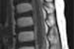
A Somatom Plus 4 Volume Zoom (Siemens Medical Solutions, Iselin, NJ) was used. The images were reconstructed to 3-mm slice thickness at 2-mm increments. The data was then volume-rendered with Siemens 3-D Virtuoso software.
"This is a very common post-processing step, which is done in almost no time," Stoeger said. "Then the data was transferred to an external high-performance workstation."
 |
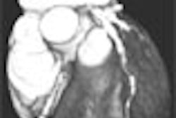
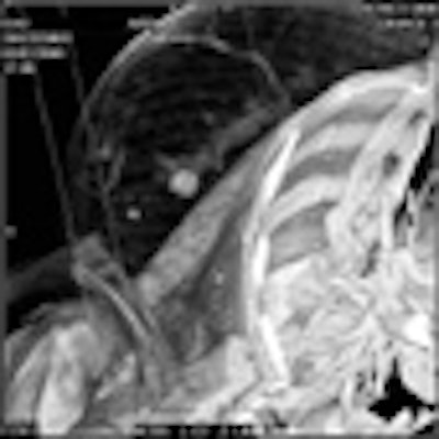
The breast images were assessed using an interactive change of rendering parameters, zooming, color encoding, and clip-plane editing in different orientations. "Clip planes can be placed in any orientation," Stoeger explained. "When placed in the oblique plane, you can acquire images that are similar to mammograms."
According to the results, breast CT found solitary solid nodules (>1.5 cm) in four patients. Single or multiple cysts (>5 mm) were found in eight patients, and macrocalcifications (>3 mm) were found in six women. Breast CT could not discern microcalcifications, which had been previously identified with mammograms, because of limited spatial resolution, Stoeger said.
The group concluded that CT was not useful for detecting microcalcifications, but that the modality may have potential for preoperative planning.
"We think that there may be a potential for this method in preoperative planning of breast disease (including the axilla) in the follow-up of certain benign masses, or even in planning radiotherapy after breast surgery," Stoeger said. "This does not mean that a CT scan should be performed for these reasons. But if these patients get a scan for other reasons, and the data exists, it could be post-processed and used for these issues instead of other time-consuming and expensive exams."
One instance where breast CT may play a role is in smokers who undergo lung cancer screening. A recent study in the June issue of Chest linked smoking to pulmonary metastasis of breast cancer. Researchers from University of California Davis Medical Center found that women whose breast cancer had spread to the lungs were nearly twice as likely to be smokers than women whose breast cancer had not spread (Chest, June 2001, Vol.119: 6, pp. 1635-1640).
By Shalmali PalAuntMinnie.com staff writer
July 3, 2001
Click here to post your comments about this story. Please include the headline of the article in your message.
Copyright © 2001 AuntMinnie.com