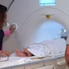
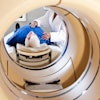
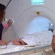
A total of 90 patients (56 males) with a mean age of 65 ± 12 years were included in the study. Participants were imaged first using a commercial chest x-ray system and then by the group's prototype dark-field chest x-ray machine, which yields both an attenuation and a dark-field image. Three independent readers assessed the conventional attenuation image and dark-field image for the presence and severity of emphysema (no emphysema, mild emphysema, moderate emphysema, severe emphysema).
According to the findings, the dark-field images showed a distinct decrease of signal strength with emphysema severity. Readers were significantly better able to identify emphysema with images from the dark-field prototype, achieving an area under the curve (AUC) of 0.85 (p < 0.05) compared with conventional images (AUC, 0.74).
In addition, while ratings of adjacent emphysema severity groups with conventional radiographs were only different for trace and mild emphysema, ratings based on images from the dark-field prototype were different for trace and mild, mild and moderate, and moderate and confluent emphysema.
"Compared to conventional chest radiography, dark-field chest radiography provides improved capability for identification and staging of emphysema," noted Theresa Urban, who will present the study.


