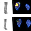
Presenter Dr. Katie Hunt of the Mayo Clinic in Rochester, MN, will share findings from a study that assessed the positive predictive values for malignancy in an MBI lexicon. The study included 550 cases of patients with a positive exam on a dual-detector MBI system performed between August 2005 and August 2007. The team tracked lesion type (mass versus nonmass uptake), distribution, intensity, and number of views on which the lesion was seen using a gamma breast imaging lexicon and correlated these results with follow-up imaging or pathology.
In 550 patients, 634 lesions were identified. Of these, 26% were malignant and 74% were benign. Hunt and colleagues found the following:
- 73% of lesions were masses.
- Most lesions (38%) were segmental in terms of distribution.
- 66% of lesions had marked intensity of uptake on MBI.
- 45% of lesions seen on four views were malignant.
Keeping these characteristics in mind can help clinicians best use MBI for diagnosing breast cancer, according to the group.
"Lesions described as masses and those with marked intensity radiotracer uptake have the highest predictive value for malignancy on MBI," Hunt and colleagues concluded.
