"The cognitive neuroscience research literature has demonstrated that resting-state BOLD fMRI is capable of measuring some features of neuronal connectivity in normal human brain," said lead author Dr. Jason Druzgal, neuroradiology fellow in the department of radiology at the University of Utah. "So, it stands to reason that we might be able to measure perturbations of connectivity that occur with many types of pathology."
A 3-tesla MRI scanner (Magnetom Trio, Siemens Healthcare, Malvern, PA) collected eight to 10 minutes of resting-state BOLD fMRI data from 24 autistic patients and 15 typically developing control patients.
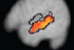
For each individual subject, the Resting State Toolkit (REST) was used to calculate the regional homogeneity value for each voxel in the brain. A group analysis then looked at every voxel in the common brain space to determine where the regional homogeneity values for the autistic patients were different from the control subjects.
The analysis showed resting-state BOLD fMRI identified differences between the brains of autistic patients and the matched control group.
"We are pretty confident in our ability to show resting-state functional MRI differences between a population of pathologic patients and their matched controls," said Druzgal, "but it is a very different question to take a single set of anonymous data and determine the population to which it belongs."
He added that more research would be necessary before these techniques can become clinically useful.