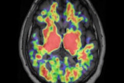
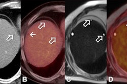
The use of DWI instead of FDG-PET/CT would mean less radiation exposure for patients.
"Currently, oncologists depend on FDG-PET/CT for the evaluation of treatment response in bone metastasis," said researcher Dr. Won-Hee Jee from the department of radiology at Seoul St. Mary's Hospital. "However, the disadvantage of FDG-PET/CT is the radiation exposure."
The study included 45 patients with pathologically confirmed soft-tissue and bone tumors. Subjects underwent 3-tesla MRI, including DWI, and whole-body FDG-PET/CT before treatment for their cancer.
The researchers calculated maximum (SUVmax) and average (SUVav) standardized uptake values and measured minimum (ADCmin) and average (ADCav) apparent diffusion coefficients of tumors on tumor regions where SUVs were obtained.
"We found that there was significant correlation between ADC with 3T DWI and SUV with PET/CT in bone and soft-tissue tumors," Jee said. "DWI [also] showed better diagnostic performance than PET/CT for diagnosing malignancy."
Because DWI can predict response in the early treatment period of bone metastasis, it would be helpful to decide whether that treatment should be continued or not, Jee added.


