(Booth 2015) Software developer MIMvista of Cleveland will highlight new features for its MIMneuro package for the quantitative analysis of PET and SPECT brain studies.
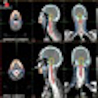
MIMneuro adds an automated workflow for epilepsy analysis, performing ictal-interictal SPECT subtractions and MR coregistration within one diagnostic reading station. MIMneuro Epilepsy Workflow is designed to lead users through the procedure step-by-step for consistent outcomes in less than five minutes.
The software's workflow automates the fusion, normalization, subtraction, and display of multiple datasets. The automated system, in turn, is designed to highlight significant foci by cluster analysis, providing radiologists and referring physicians with an improved image of the epileptic focus.

MIMvista also will display its MIM Storage Server in Chicago. MIM Storage Server provides department staff and referring physicians with access to image data, offering archive searching and data transfer with automated study routing and image retrieval capabilities.
MIM Storage Server also allows for storage of specific formats, including RT Dose, RT Plan, and RT Struct. Scalable security options are provided using LDAP/Active Directory protocols, and storage can be expanded with readily available hardware. MIM Storage Server is FDA-cleared and is currently available for use in radiology, nuclear medicine, radiation oncology, and other clinical applications.
MIMvista also will offer its MIMcontouring tools for advanced serial exam review and therapy response assessment. Multiple modalities such as CT, MR, PET, and SPECT can be automatically linked or fused for comparison. Triangulation of the fused or linked exams allows corresponding abnormalities to be tracked between time points.
PET exams coregistered to the CT also can be aligned with enhanced accuracy across time points, utilizing the same deformation from the CT. This registration method also can be used to segment tumors on a new CT exam through a deformable recontouring technique.
Tumors on PET exams can be segmented accurately in seconds using PET Edge. Tumor statistics, such as volume, SUVmax, SUVmean, and response evaluation criteria in solid tumors (RECIST), and screen captures can be added to customizable reports or saved to a MIMviewer DICOM CD for referring physician review.
MIMvista's Mobile MIM Pro software also debuts at RSNA 2008. It features remote image viewing on Apple's iPhone or iPod Touch with multitouch interface, including zoom, fusion, blending, window/level, standardized uptake values, measurement tools, and contour review. Key features are multiplanar reconstruction, contour review, and data encryption, which ensures patient privacy. Mobile MIM Pro is pending FDA clearance.
With applications in teleradiology, healthcare services marketing, and referring physician communication and collaboration, Mobile MIM Pro will be available worldwide in 2009.
MIMvista also will show its MIMcontouring VoxAlign deformation engine, designed to allow for atlas-based segmentation and recontouring for adaptive therapy and gated studies. Atlas-based segmentation automatically generates entire structure sets, including target volumes, dramatically reducing time for initial contour generation.
Entire gated CT studies can be contoured automatically by deforming contours from a single phase to match the anatomy on all phases. Deformable coregistration also is designed to correct for nonrigid differences in alignment, allowing PET/CT images to be aligned with greater accuracy to the planning CT, even when the PET/CT was not obtained in treatment position.
The MIMcontouring VoxAlign deformation engine currently is available worldwide.