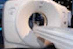
One of the most common indications for CT imaging of the head is the probability of an acute cerebral ischemia, whereby the patient usually undergoes a CT perfusion study supplemented by an additional 3D CTA for vascular evaluation, Dr. Elyas Ghariq, from Leiden University Medical Center, told AuntMinnie.com.
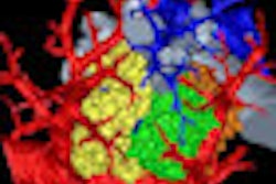
With the introduction of the 320-slice CT scanner came the possibility of depicting the whole head in a single rotation. With this came the possibility of acquiring so-called "whole-head" CT perfusion studies, and the option to reconstruct dynamic CT angiography exams (4D CTA) from CT perfusion data.
CT perfusion studies on a 320-detector-row scanner (Aquilion One, Toshiba Medical Systems) usually require less radiation, Ghariq said. However, early studies showed that due to the lower dose and broader conebeam angle of 320-detector-row CT, 4D CTAs reconstructed from these data had higher noise levels and less vascular quality compared to additional 3D CTAs, especially in the small arteries.
Also, 4D CTA usually requires an optimized workstation for 320-slice CT with a built-in subtraction tool to mask bone and other nonvascular structures. However, a recent study described the derivative-based method of obtaining vascular images from 64-slice CT perfusion data; it showed that when using the method, arterial quality is similar compared to standard CTA on a 64-detector-row scanner. The derivative-based method enhances structures to show density changes in time (vascular structures) while masking nonvascular areas with static Hounsfield values.
The researchers investigated whether the new technique can reconstruct CTAs with improved arterial quality from 320-slice CT perfusion studies, and whether the improved quality is comparable to the arterial quality of the additional 3D CTAs acquired on 64-slice CT scanners. If the quality is high enough, the additional 3D CTA scans can be skipped, Ghariq said.
The study found significant quality improvement in all large extradural and intradural arteries, especially in small arteries, when derivative-based reconstructed 4D CTAs were compared with 4D CTAs reconstructed using the built-in subtraction tool on an optimized workstation for 320-slice CT data.
There were no significant differences in arterial image quality between 64-slice 3D CTAs and derivative-based reconstructed 4D CTA. The study showed that 4D CTA derived from existing 320-slice CTP data has the potential to replace CTA, when both CTA and CT perfusion are required, reducing radiation dose to the patient.