Advanced visualization displays certainly have a role in communicating information about cases to referring clinicians. Yet there remains "an unmet need to render DICOM images as 3D-printed models capable of providing both tactile feedback and tangible depth information of anatomic and pathologic states," said course instructor Dr. Frank Rybicki, PhD. Rybicki is chair of radiology at the University of Ottawa and editor in chief of the new peer-reviewed Springer journal 3D Printing in Medicine.
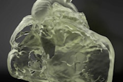
A growing body of data supports the use of models to capture complex anatomy, such as congenital heart disease requiring surgery, and 3D-printed models are being rapidly embraced in cardiovascular care, he said.
The lecture will summarize studies to date supporting the benefits of cardiovascular 3D printing, and look at the projected growth of 3D-printed models generated mostly from DICOM CT and MR images for planning, intervention, and fabrication of implants. Rybicki will also discuss the most exciting applications in cardiovascular 3D printing.
"For newborns with congenital heart disease, there is already a large body of evidence that 3D models aid in surgical planning, even in comparison with so-called '3D visualization,' which includes volume rendering as well as multilayer reformatting of DICOM images," he wrote. "In addition, there is a growing use of 3D printing for patients with valve disease, in particular for planning less invasive intervention for pathologies of the aortic valve and the mitral valve."