Lobar Swyer-James Syndrome:
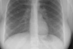
(Click in small images to view the larger radiographs)The patient shown below was a middle aged male with a long history of
obstructive airway disease on pulmonary function testing. His PA chest
radiograph demonstrated hyperlucency in the right lower lung.

A CT scan of the chest was performed and revealed decreased attenuation
within the right middle and lower lobes, with a decreased number of vascular
markings. Bronchiectatic changes were also noted in the right lower lobe
(red arrows). Bronchoscopy was negative, and the findings were felt to
most likely be related to a lobar Swyer-James syndrome.