CHICAGO - Intrarenal color Doppler ultrasonography may be the answer to finding a noninvasive and accurate method to diagnose renal artery stenosis (RAS). A soon-to-be-published study lays down parameters through which RAS detection is improved to nearly 100% through intrarenal color Doppler US, giving it an edge over captopril scintigraphy and conventional Doppler of the main artery.
The study, done at the B.P. Koirala Institute of Health Sciences in Dharan, Nepal, and presented at the RSNA meeting this week, finds "Intrarenal color Doppler US with additional sampling from the upper and lower pole is an accurate method for screening and diagnosing significant RAS, including stenosis of accessory renal artery." Significant RAS was stenosis of more than 60%.
Captopril scintigraphy and intrarenal color Doppler ultrasonography were done in 26 patients with suspected RAS to determine which was the more accurate screening tool. The results were interpreted independently by different operators.
"Clear selection of patients for DSA (digital subtraction angiography) and therapeutic angiography are not available. So this prospective study was carried out to assess the accuracy of both methods," said Dr Raj Rauniyar, additional professor, department of radiodiagnosis at B.P. Koirala Institute of Health Sciences.
DSA was used as the gold standard, and patients were grouped into four categories based on severity. Group 1 included patients with 0 to 59% stenosis, group 2 patients with 60% to 95% stenosis, group 3 patients with 95% to 99% stenosis, and group 4 patients with 100% occlusion.
According to the results, captopril scintigraphy had a sensitivity of 75%. It failed to detect two instances of accessory artery stenosis.
On the other hand, using parameters such as acceleration time (AT), acceleration time ratio (ATR), acceleration index (AI), and characteristic of spectral trace (parvus tardus), Doppler had an overall detection of nearly 100% with intrarenal branch arteries.
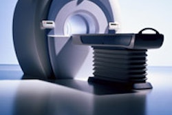
The color Doppler US was done using an ATL Ultramark 9 HDI system (Philips Medical Systems, Bothell, WA) with 3.5-MHz transducers.
A limitation of intrarenal color Doppler US was that it was not able to differentiate between group 3 and group 4 patients, and diagnosis in these cases was subjective, Rauniyar said. But since patient management does not vary and calls for DSA for both groups, the researchers concluded that intrarenal color US is superior to captopril scintigraphy and conventional Doppler of the main artery.
Conventional Doppler of the main artery has the disadvantage of being time-consuming and operator-dependent. In the patient group, the study of the main renal artery was time-limited to 15 minutes for each artery. Visualization was poor and success rate was 55.81%, according to Rauniyar.
But with color Doppler US, almost all cases were diagnosed. "Compared to previous studies, results were much improved," Rauniyar said. "Color Doppler is accurate, but the clear diagnostic parameters for using it are not available."
The secondary goal of the study was to determine the most reliable Doppler parameters, and the research findings indicate that they are AT, ATR, AI, and parvus tardus.
By N. Shivapriya
AuntMinnie.com staff writer
December 2, 2004
Related Reading
Study finds link between RAS and cardiovascular disease risk, April 19, 2004
Brazilian sonologists offer technique for suspected RAS, June 6, 2003
Extent of coronary disease predicts presence of renal artery stenosis, November 14, 2002
Copyright © 2004 AuntMinnie.com