CHICAGO - The use of advanced radiological techniques already make it possible to perform a forensic autopsy, according to European researchers, although they continue to perform studies needed to convince pathologists and courts that "virtopsy" can become an accepted part of criminal procedures.
"We have performed 100 cases of virtual autopsy as we develop this procedure," said Dr. Michael Thali, a board-certified forensic pathologist and project manager for virtual autopsy at the Institute of Forensic Medicine at the University of Bern, Switzerland. "The virtual autopsy does not destroy evidence -- which may be damaged during a classical autopsy. It can also be used in cultures and situations where autopsy is not tolerated by religion. We think this will change the field of forensic autopsy.
"It is the autopsy of the future," he said. "We will be able to perform autopsies without a scalpel."
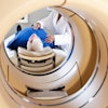
In a presentation at the 2003 RSNA meeting, Thali demonstrated how the MRI head scan of a blunt trauma patient matched the classical autopsy results. He also showed how virtopsy could reveal subtleties in anatomy that make it possible to see the track of a bullet through a victim's brain.
Body surface mapping techniques, computed tomography and magnetic resonance imaging contribute to the "virtual knife" that pathologists can use to observe the cause of death.
Thali said that getting virtopsy approved for use in the courtroom will require at least another decade of comparison studies. In each of the 100 cases performed so far, a classical autopsy has followed for use in criminal cases.
"I think in countries like the United States, the court system is even more conservative and may take a longer time," he said. "We need more validation before we will be ready to go into the court room."
Dr. Edmund Donoghue, chief medical examiner for Cook County, Ill., which includes the city of Chicago, said pathologists have been using imaging techniques to assist in autopsies for years. However, he said, the use virtopsy results as evidence in a trial is a long way off.
"It is interesting that the head wound case involved an entrance and exit wound," he said, "because that case doesn't involve recovering the bullet -- evidence -- from the victim. That is something that cannot be achieved virtually."
The equipment required to perform a virtual autopsy would strain the budgets of medical examiners, Donoghue said, and the MRI time required would tax the ability of his office to keep up. "We perform 17 autopsies a day, and to take three hours to due a virtual autopsy would not be practical," he said.
Dr. Anna Graham, professor of pathology at the University of Arizona Health Sciences Center, said the use of virtopsy might be best employed as a teaching tool for pathologists, especially in examining disease states.
Thali virtopsy is a natural result of merging radiological techniques into forensic work. In their virtopsy work, Thali and colleagues use both multi-slice CT images obtained on a GE Lightspeed QX/i unit, and MR images from a GE 1.5-T Signa Echospeed Horizon, version 5.8.
Postmortem diagnostic imaging is generally underutilized, Thali said. The cost of and limited access to equipment, coupled with lack of awareness of its potential, has slowed the development of the procedure.
He noted that MR has long been used in paleoimaging to examine mummies without damaging them. He also said that micro-imaging with CT and MRI will add a new dimension to histopathology, perhaps making image-guided virtual histology a reality.
By Edward SusmanAuntMinnie.com contributing writer
December 4, 2003
Copyright © 2003 AuntMinnie.com