CHICAGO - Using a gantry rotation in the coronal plane to image only the breast, physicists from the University of California believe they have developed a dose-efficient technique comparable to two-view mammography.
Dr. John Boone from UC Davis presented the results of his work with Dr. Thomas Nelson, from UC San Diego at the RSNA meeting on Tuesday.
"Over the last 20 years, CT has been largely dismissed with the thought that the radiation dose would be too high for breast screening,” Boone said. With traditional mammography, the breast must be scanned axially in order to penetrate the thorax. In addition, there is a greater radiation dose to the surrounding tissues, a larger field of view is required, and cardiac motion is an issue.
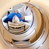
In comparison, dedicated breast CT scans can be done in the coronal plane with the breast hanging pendulantly, as in a biopsy, Boone said. A smaller field of view and lower radiation dose are two other advantages of this technique, he added.
Boone and Nelson used Monte Carlo techniques to assess radiation dose to the breast. A total of 107x-ray photons at energies from 10 keV to 140 keV, at 1 keV intervals, were followed. A polyenergetic tungsten-anode x-ray spectral model was used for dose assessment.
The photon fluence to the detector equivalent to that of a high dose abdominal CT scan was assumed. Dose to the breast at constant signal-to-noise ratio was computed from 30 kVp to 140 kVp in 5 kVp intervals.
For subjective assessment of image quality, a cadaver breast was scanned at 80 kVp, on a GE Medical Systems LightSpeed, using 1 mm contiguous slices and 300 µm x 300 µm x 1.0 mm voxels.
According to the results, at the center of 10 cm diameter breasts, CTDI values were determined as: 0.58, 2.6, 6.1, 10.6, 15.8, and 21.5 mGy/100 mAs at 40, 60, 80, 100, 120, and 140 kVp, respectively.
The dose for a 10 cm diameter breast at 80 kVp was 3.87 mGy, Boone reported, making it comparable to two-view mammography. At 100 kVp, the dose was 3.56 mGy; at 120 kVp, the dose was at 3.47 mGy. Radiation doses were unacceptably high below 40 kVp, Boone reported.
In a second presentation, Nelson discussed how this CT technique played out on a cadaver breasts. They used a volumetric review of high-resolution breast CT image data to visualize internal architecture.
“Calcifications, while important, are not the only issue that we need to deal with,” Nelson said. “In the superposition [with traditional mammography], it can be difficult to evaluation structural complexity.”
Cadaver breasts were scanned at 80-120 kVp using 1 mm slices, using 300 µm2 x 1.0 mm voxels through the entire breast, yielding a volume data set at 5123 voxels. Interactive visualization and review of multiplanar slices and volume-rendered images was done with a dedicated volume-rendering card. Glandular patterns, optimal slice orientation, and overall tissue architecture was evaluated.
According to the results, images of the cadaver breast show impressive detail of the parenchyma and ductal structure, Nelson said.
“Because this system is interactive, we can slide the slice around and view the data however we like. The information is readily accessible,” he said.
Boone and Nelson said future research in this area will focus on refining the technique so that it can visualize all the way to the chest wall. Cost and patient throughput are other issues that will need to be addressed.
Finally, the duo expect that they will have to build a dedicated breast CT system as current systems will not rotate in a horizontal plane, Boone said.
By Shalmali PalAuntMinnie.com staff writer
November 28, 2000
Copyright © 2000 AuntMinnie.com
Click here to view the rest of AuntMinnie’s coverage of the 2000 RSNA conference.
Click here to post your comments about this story.