SALT LAKE CITY – Interacting with physicians from around the world is one of the main reasons why radiologists like working at the Olympic Polyclinic. In most cases, the coaches and team doctors review the MRI with the radiologist.
Sometimes there is a language barrier, however, and no interpreter was available in this case. Fortunately, the English-speaking radiologist and the foreign team doctor soon found out they had an important word in common: syndesmosis. Much hand waving and pointing at images followed, and the two were able to discuss this syndesmosis sprain.
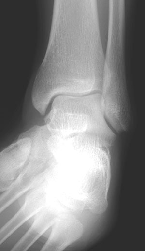 |
| Mortise view of the ankle; width of the mortise is normal. |
 |
 |
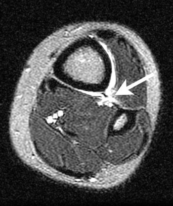
| Axial FSEIR at the tibial plafond shows rupture of the anterior and posterior tibiofibular ligaments, and the interosseous membrane (arrow). |
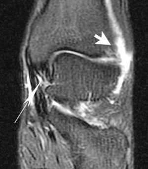 |
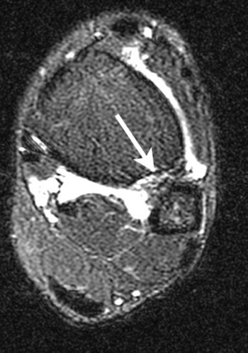
| Coronal FSEIR shows fluid (short arrow) at the anterolateral aspect of the ankle related to the syndesmosis sprain. It also demonstrates sprain of the deltoid ligament (long arrow). |
By Dr. Julia Crim
AuntMinnie.com contributing writer
February 20, 2002
Thursday: Midfoot fracture dislocation.
Dr. Crim is chief of musculoskeletal imaging at the Olympic Polyclinic.
Related Reading
Scenes from the Polyclinic: A case study in the ACL-deficient knee, February 19, 2002
Medical imaging goes for gold at Olympic Polyclinic, February 8, 2002
Copyright © 2002 AuntMinnie.com


