A traditional imaging workup for colorectal carcinoma liver metastases (CCLM) calls for a CT scan followed by ultrasound and MR for disease staging and patient management. Although PET has demonstrated superior sensitivity for cancer detection in numerous studies, few facilities use the technology as a first-line modality. A research team in Ireland used the five-step process of evidence-based medicine to successfully implement new protocols for CCLM that utilize PET as the primary imaging modality.
"The underlying theme to our research is how one can actually apply evidence-based medicine to your practice," said Dr. John Sheehan, a radiologist at St. Vincent's University Hospital Blackrock Clinic PET Center in Dublin, Ireland, who presented the results of his group's retrospective analysis at the European Congress of Radiology (ECR) in Vienna, Austria, earlier this month.
Sheehan said that evidence-based practice calls for clinicians to take five steps:
- Ask a focused clinical question.
- Search for best current evidence.
- Appraise validity and strength of evidence.
- Apply to patients and practice.
- Evaluate performance.
Steps 1-3
At Sheehan's facility, the imaging protocol for CCLM started with a CT exam of the abdomen and pelvis taken by a referring hospital. The facility would then conduct a CT scan of the patient's brain, thorax, abdomen, and pelvis. If necessary, the staff would order ultrasound and MR exams. The researchers focused their efforts on determining if their CCLM imaging protocol was utilizing the best technology for staging CCLM.
The group conducted a meta-analysis of published medical literature in an effort to uncover the most sensitive modality for staging CCLM.
"What we found was that the strongest, most sensitive modality was PET, with a sensitivity of 90%," Sheehan said.
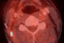
From July 2002 through December 2003, the researchers examined 40 consecutive patients (25 male, 15 female) with colorectal carcinoma who had a PET study prior to liver resection. Radiologists at the facility reviewed the PET images, and compared them with each patient's CT and MR exams.
"The patient records were then reviewed to establish the surgical intentions before and after the PET study," Sheehan said.
Of the 40 patients, the radiologists determined 38 (95%) exhibited an abnormality, while two patients (5%) were reported as normal. The cohort consigned to the abnormal category was further refined based on hepatic metastasis (30 patients or 75%) or extrahepatic abnormalities (21 patients or 53%). Sheehan said that 18 patients (47%) were judged suitable for hepatic resection.
Steps 4-5
The researchers reported that patient management was altered in 35% of the cases (15 patients). Sheehan said that 11 of the patients were upstaged to unresectable, two were downstaged to resectable, and one was upstaged for resection. A total of 18 patients underwent surgical resection, and there was concordance in 16 (89%) of the cases. Five of the patients with limited extrahepatic disease on PET underwent laparotomy and CCLM resection, Sheehan noted.
"The discordances in patient management were due to one patient with a tumor adherent to the diaphragm and one young patient with an equivocal PET result that were unresectable at laparotomy," he said.
The team found that the diagnostic impact of using PET or conventional imaging was even more significant, with a total discordance of 64% between PET and CT, MRI, and ultrasound. In the cases of hepatic metastasis, there was discordance in 15 patients (40%), and in the cases with extrahepatic abnormalities, there was discordance in nine patients (24%). Sheehan explained that the majority (70%) of the total discordance was due to tumor upstaging due to PET imaging.
The researchers reviewed their analysis, and concurred that the use of PET for staging CCLM had a major impact on patient care (64% diagnostically, 35% on patient management) compared with the previous imaging protocol of CT with MR and ultrasound.
"Our practice now is to review the referring CT scan, and simply go on and perform a PET scan," Sheehan said.
By Jonathan S. Batchelor
AuntMinnie.com staff writer
March 18, 2005
Related Reading
Colorectal cancer survival lower in Europe than in U.S., February 16, 2005
Coffee may reduce risk of liver cancer, February 16, 2005
Red meat linked to one type of colon cancer, February 3, 2005
CT tracks renal cancer recurrence after surgery, January 20, 2005
PET/CT improves detection of metastatic colorectal cancer, December 16, 2004
Copyright © 2005 AuntMinnie.com