Fibroblast activation protein inhibitor (FAPI)-PET diagnostic cancer imaging may be performed reliably as early as 10 minutes after radiotracer injection -- a significant reduction of the typical one-hour wait period for tracers such as FDG-PET, European researchers have found.
A team led by Dr. Frederik Giesel of University Hospital Dusseldorf in Germany compared incubation times of gallium-68 (Ga-68) FAPI-46 in different cancers at three time points after patients received the radiotracer, including after the conventional one-hour wait. The group learned that FAPI-46 is highly reliable as early as 10 minutes after injection. The results of the study were published November 10 in the Journal of Nuclear Medicine.
"This could have important implications for improving workflow and decreasing wait times in nuclear medicine departments compared with more traditional PET agents such as FDG-PET," the authors wrote.
Fibroblast activation protein (FAP) is overexpressed in cancer, and researchers suspect it is an essential component driving the growth of tumors. Experimental FAP-targeted molecular imaging agents, such as FAPI-04 and FAPI-46, have shown promising results in tumor diagnosis.
By convention, most molecular imaging tracers such as FDG are injected one hour prior to PET scans to allow time for uptake of the tracer by tumors. Yet there are conflicting reports about optimal incubation times for these new FAPI agents, with no conclusive data published, the authors noted.
To address the knowledge gap, Giesel and colleagues retrospectively analyzed results from 43 patients with diverse cancer diagnoses who each underwent Ga-68 FAPI-46-PET/CT scans at 10 minutes, one hour, and three hours after tracer injection. They determined the tracer uptake in patient tumors based on standardized uptake values (SUVmean and SUVmax) and tumor-to-background ratios (TBR).
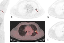
 A case example of a 63-year-old patient with esophageal cancer. FAPI-46 PET/CT was performed for irradiation planning before definitive radiochemotherapy. FAPI-46 PET was performed 10 minutes, one hour, and three hours after injection. Image courtesy of the Journal of Nuclear Medicine.
The FAPI-PET scans revealed 171 lesions in the 43 patients. Comparing all lesions at different time points, the mean SUVmax was maximal at 10 minutes (8.2) and declined slightly at one hour (8.15) and at three hours (7.6) after tracer administration, the team found.
Additionally, the mean SUVmax had a similar pattern in primary lesions, lymph node metastases, and distant metastases. TBRs also showed nonsignificant differences at the three times.
Ultimately, although this was a small study, due to the similar detection rate between 10 minutes and one hour, the authors suggest 10 to 20 minutes after injection is the best time point for diagnostic PET imaging acquisition.
"Confirmation of these findings in a larger cohort is underway," they wrote.