SPECT/CT imaging appears more accurate than standard contrast-enhanced CT exams for diagnosing benign versus malignant kidney tumors, according to a study published June 30 in the Journal of Nuclear Medicine.
A U.S. group in Missouri analyzed scans from patients with biopsy-proven renal masses who underwent both SPECT/CT with technetium-99m sestamibi (Tc-99m) and contrast-enhanced CT. The team found SPECT/CT better distinguished between benign and malignant tumors.
"This can help in potentially avoiding overtreatment in patients with relatively indolent tumors, while appropriately managing the more aggressive malignancies," wrote corresponding author Dr. Ashwin Singh Parihar, a postdoctoral radiology research associate at Washington University in St. Louis.
The American Cancer Society estimated that 30,520 deaths (20,420 men and 10,100 women) from liver cancer will occur in the U.S. this year. Most renal masses are malignant, with renal cell carcinoma (RCC) forming the most common type, but up to 20% may be benign.
Differentiating between these tumors is a diagnostic challenge, as conventional imaging with CT or MRI cannot reliably differentiate the benign and indolent tumors from the more aggressive ones, the study authors stated. This can lead to overtreatment in patients with relatively indolent tumors, they wrote.
Moreover, while Tc-99m sestamibi SPECT/CT is not yet currently recommended as an appropriate imaging tool for indeterminate renal masses by the American College of Radiology (ACR), this is largely due to the limited patient numbers in studies supporting its use, the Parihar and colleagues added.
Thus, the group sought to provide additional evidence on the diagnostic accuracy of the approach over contrast-enhanced CT.
The researchers analyzed imaging and clinical records of patients who underwent Tc-99m sestamibi SPECT/CT for clinical workup of solid renal masses from September 2018 to October 2021 at the school's Mallinckrodt Institute of Radiology. They identified 42 patients. A histopathologic diagnosis (from biopsies or after surgical excision) was available in 27 of these patients, of whom 20 also had contrast-enhanced CT exams.
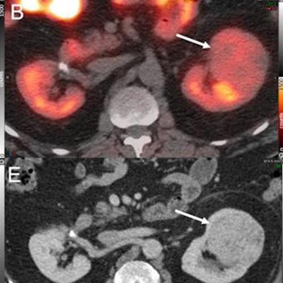
 A 67-year-old man with previously diagnosed right renal cell carcinoma (RCC), post right partial nephrectomy presented with an enlarging left renal mass. Tc-99m sestamibi axial SPECT (A), and fused SPECT/CT (axial = B, coronal = C) images show a mass in the superior pole of the left kidney with its inferior part showing tracer avidity (thin arrow) and the superior part showing relative photopenia (thick arrow). It was diagnosed as a likely collision tumor on SPECT/CT with the inferior, tracer-avid region likely representing an oncocytic tumor and the superior, photopenic region likely representing a concerning RCC. Contrast-enhanced CT images (noncontrast = D, postcontrast nephrogenic phase axial = E, coronal = F) showed a solid heterogeneously enhancing left renal mass likely representing oncocytoma. The patient underwent left partial robot-assisted laparoscopic nephrectomy, and histopathology showed a collision tumor composed of clear cell RCC (rhabdoid differentiation, WHO/ISUP grade 4) and an eosinophilic variant of chromophobe RCC. Image courtesy of the Journal of Nuclear Medicine.
Based on the biopsies, the researchers split the tumors into 19 "malignant/concerning" tumors (52.8%) and 17 "benign/nonconcerning" (47.2%) tumors. They then performed visual interpretations of the two imaging approaches to calculate their sensitivity and specificity for characterizing the tumors in each group.
The sensitivity and specificity of SPECT/CT for diagnosing nonconcerning lesions were 66.7% and 89.5%, respectively, compared with 10% and 75% for contrast-enhanced CT, according to the analysis. In addition, SPECT/CT correctly classified 80% of the RCC lesions, compared with 33.3% on contrast-enhanced CT.
"The incremental value of SPECT/CT in detecting the oncocytic lesions [benign] is clinically relevant and can potentially avoid overtreatment in these patients," the researchers wrote.
The authors noted that the literature is sparse in this space in terms of original articles describing findings in unique populations, yet the stringent inclusion criteria with histopathology as the reference standard helped form a robust comparison between the two approaches.
"We provide additional evidence on the utility of Tc-99m sestamibi SPECT/CT, especially in patients with an inconclusive [contrast-enhanced] CT for differentiating the malignant/aggressive renal tumors from the benign/indolent ones," Parihar and colleagues concluded.