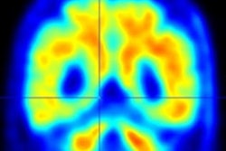
Researchers have developed a new model for diagnosing the severity of Alzheimer's disease in patients based largely on the spatial topography of brain PET imaging, according to a study published April 25 in Nature Aging.
An international group led by Canadian researchers applied topographical data from tau PET scans to an anatomical framework called Braak staging in a group of patients with Alzheimer's disease. They found that their PET-based Braak staging model's stages were associated with cognitive dysfunction and severity of dementia.
Ultimately, the model could help clinicians monitor patients as Alzheimer's disease progresses, from early stages to clinical dementia, according to the group.
"Using PET-based Braak staging provides an opportunity to study the dynamics of [Alzheimer's disease] biomarker changes in living individuals," wrote corresponding authors Dr. Joseph Therriault and Dr. Pedro Rosa-Neto of McGill University in Montreal, Quebec, Canada.
Neurofibrillary tangles of tau protein in the brain are a hallmark of Alzheimer's disease. Braak staging was developed in the 1990s through postmortem analysis of these tangles in brain samples.
The Braak system stages patients with Alzheimer's disease according to tau aggregation in the transentorhinal cortex (stage I), entorhinal cortex and hippocampus (stage II), inferior temporal neocortex (stage III), association cortices (stages IV and V), and primary sensory cortices (stage VI).
Although Braak staging is part of the gold standard diagnostic workup for Alzheimer's disease at autopsy, it remains a largely histopathological construct and has yet to be integrated into the definition of the disease in patients, the authors wrote.
In this study, the group used data from patients who underwent previous imaging using an experimental tau PET radiotracer called F-18 MK-6240, which has shown a high affinity for binding to tau protein, especially over time.
The group hypothesized that tau accumulations on PET scans would follow the hierarchical pattern established by Braak staging and that the model could indicate memory impairment in early and late stages of Alzheimer's disease in patients.
The researchers identified 179 cognitively unimpaired (CU) older adults, 80 individuals with mild cognitive impairment (MCI), and 65 individuals with Alzheimer's disease clinical dementia from previous studies. The mean age of patients was 70.05 years old, and 60% were women.
First, the researchers assigned PET-based Braak stages to individual patients ranging from 0 (no detectable tau-PET abnormality) to VI (tau-PET abnormality extending to primary sensory areas) based on the accumulations of tau observed on PET scans. Next, they assessed the relationship between PET-based Braak stages and clinical dementia severity according to the Clinical Dementia Rating (CDR), an interview tool clinicians use to apply scores to cognitive function.
Analysis revealed that PET-based Braak staging tracked Alzheimer's disease symptoms from memory impairment to dementia, the authors wrote.
When they assessed the relationship between CDR and PET-based Braak stage, they observed that Braak stages 0, I, and II were compatible with normal cognition.
Braak stage III and IV were most frequently associated with a CDR of 0.5, which is commonly observed in individuals with MCI. Some individuals at PET-based Braak stage V had a CDR of 1, indicating mild dementia, whereas PET-based Braak stage VI was associated with mild and moderate dementia, according to the findings.
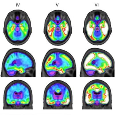
 Average F-18 MK-6240 SUVRs across the whole brain for all participants, grouped according to PET-based Braak staging. Individuals at stage 0 did not have detectable tau abnormality in regions comprising any Braak stage. At stage I, tau abnormality is confined to the transentorhinal cortex. Braak stages V and VI are characterized by high magnitude of tau pathology, with stage VI extending to primary sensory cortices. Image courtesy of Nature Aging.
In addition, in a subsample of individuals with autosomal dominant Alzheimer's disease, the researchers found the PET-based Braak stage increased sequentially up to the estimated onset of symptoms.
"Although these results require replication in larger cohorts, symptom onset took place approximately between Braak stages II and IV, with increasing PET-based Braak stage associated with the continued worsening of cognitive impairment," the authors wrote.
The authors noted that like other staging systems in medicine, it is important to emphasize that such disease staging in Alzheimer's disease patients is not intended to replace assessment of symptoms. Rather, it can be used in tandem to provide complementary information about the disease course, especially at early stages, they wrote.
"Our results support PET-based Braak staging as a framework to model the natural history of [Alzheimer's disease], as well as monitor [Alzheimer's disease] severity from presymptomatic to clinical dementia phases," the group concluded.